 Skip to content
Media
{"description":"This vibrant corporate film reveals GDT’s core philosophy: innovation, transparency, and global partnership. It intercuts close‑ups of clinician workshops in Rome, São Paulo, and Hong Kong with slow‑motion factory shots of titanium shavings dancing to an original symphonic score. Dual Hebrew‑English narration notes that the company reinvests 15 % of revenue in R\u0026D, covering Nano‑Etch surfaces and 10‑million‑cycle fatigue tests. Dentists praise the free G‑Academy virtual courses and subsidized hands‑on sessions. Humanitarian clips show GDT teams volunteering in Uganda, with patients beaming after fixed restorations. The final message—“The vision behind every smile is trust”—appears over a Tel Aviv sunset, inviting new partners to join the dental revolution.","media_image":"https:\/\/i.ytimg.com\/vi\/lDFx0wGQtaI\/hq720.jpg?sqp=-oaymwEnCNAFEJQDSFryq4qpAxkIARUAAIhCGAHYAQHiAQoIGBACGAY4AUAB\u0026rs=AOn4CLCQyA_TiGW2ZjHV-5-LnUDejUQ7pw","media_link":"https:\/\/www.youtube.com\/watch?v=lDFx0wGQtaI\u0026pp=ygUzVGhlIFZpc2lvbiBCZWhpbmQgRXZlcnkgU21pbGUgLSBHRFQgRGVudGFsIEltcGxhbnRz","media_page_content":{"type":"root","children":[{"type":"paragraph","children":[{"type":"text","value":"This vibrant corporate film reveals GDT’s core philosophy: innovation, transparency, and global partnership. It intercuts close‑ups of clinician workshops in Rome, São Paulo, and Hong Kong with slow‑motion factory shots of titanium shavings dancing to an original symphonic score. Dual Hebrew‑English narration notes that the company reinvests 15 % of revenue in R\u0026D, covering Nano‑Etch surfaces and 10‑million‑cycle fatigue tests. Dentists praise the free G‑Academy virtual courses and subsidized hands‑on sessions. Humanitarian clips show GDT teams volunteering in Uganda, with patients beaming after fixed restorations. The final message—“The vision behind every smile is trust”—appears over a Tel Aviv sunset, inviting new partners to join the dental revolution."}]}]},"title":" The Vision Behind Every Smile - GDT Dental Implants"}
The Vision Behind Every Smile - GDT Dental Implants
This vibrant corporate film reveals GDT’s core philosophy: innovation, transparency, and global partnership. It intercuts close‑ups of clinician workshops in Rome, São Paulo, and Hong Kong with slow‑motion factory shots of titanium shavings dancing to an original symphonic score. Dual Hebrew‑English narration notes that the company reinvests 15 % of revenue in R&D, covering Nano‑Etch surfaces ...
Unboxing GDT
{"description":"The GDT Hemosponge 100% Pure Collagen Absorbable Sponge is a gamma-sterilized collagen matrix made from 100% Pure Type 1 Bovine Collagen. Each pack includes 10 sponges for maximum safety. Measuring Ø10.0mm by 20.0mm, it excels in healing with bioabsorbable collagen, ensuring superior sterility, prolonged effectiveness, and exceptional hemostatic control.\n","media_image":"https:\/\/i.ytimg.com\/vi\/qv4CT-JsXUw\/hq720.jpg?sqp=-oaymwFBCNAFEJQDSFryq4qpAzMIARUAAIhCGAHYAQHiAQoIGBACGAY4AUAB8AEB-AH-CYAC0AWKAgwIABABGBkgaihyMA8=\u0026rs=AOn4CLCBvLeT2ZLiPiRxqDs0lVmaBccMOQ","media_link":"https:\/\/www.youtube.com\/watch?v=qv4CT-JsXUw","media_page_content":{"type":"root","children":[{"type":"paragraph","children":[{"type":"text","value":"The GDT Hemosponge 100% Pure Collagen Absorbable Sponge is a gamma-sterilized collagen matrix made from 100% Pure Type 1 Bovine Collagen. Each pack includes 10 sponges for maximum safety. Measuring Ø10.0mm by 20.0mm, it excels in healing with bioabsorbable collagen, ensuring superior sterility, prolonged effectiveness, and exceptional hemostatic control.\n\nDesigned for versatility, GDT Hemosponge supports guided bone regeneration, preventing migration for successful outcomes. Its durability minimizes alveolar ridge atrophy, reinforcing structural support.\n\n"}]}]},"title":"Unboxing GDT Hemosponge -100% Pure Collagene"}

Unboxing GDT Hemosponge -100% Pure Collagene
The GDT Hemosponge 100% Pure Collagen Absorbable Sponge is a gamma-sterilized collagen matrix made from 100% Pure Type 1 Bovine Collagen. Each pack includes 10 sponges for maximum safety. Measuring Ø10.0mm by 20.0mm, it excels in healing with bioabsorbable collagen, ensuring superior sterility, prolonged effectiveness, and exceptional hemostatic control.
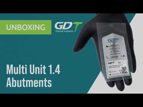
{"description":"An aluminum packet opens to four Multi‑Unit 1.4 mm abutments in varying collar heights. The TiAlN‑coated screw supports 30 N cm torque. FEM analysis shows even stress distribution. A clinical clip installs them in an upper All‑on‑4; a Sheffield test confirms passive fit of the provisional PMMA bridge. A cost slide shows 20 % savings over imported titanium bases. A green color band ensures quick OR identification. The product is CE‑marked and listed in the French reimbursement catalog.","media_image":"https:\/\/img.youtube.com\/vi\/tuuUlir2VLk\/hqdefault.jpg","media_link":"https:\/\/www.youtube.com\/watch?v=tuuUlir2VLk","media_page_content":{"type":"root","children":[{"type":"paragraph","children":[{"type":"text","value":"An aluminum packet opens to four Multi‑Unit 1.4 mm abutments in varying collar heights. The TiAlN‑coated screw supports 30 N cm torque. FEM analysis shows even stress distribution. A clinical clip installs them in an upper All‑on‑4; a Sheffield test confirms passive fit of the provisional PMMA bridge. A cost slide shows 20 % savings over imported titanium bases. A green color band ensures quick OR identification. The product is CE‑marked and listed in the French reimbursement catalog."}]}]},"title":"Unboxing GDT Multi Unit 1.4mm Abutments"}
Unboxing GDT Multi Unit 1.4mm Abutments
An aluminum packet opens to four Multi‑Unit 1.4 mm abutments in varying collar heights. The TiAlN‑coated screw supports 30 N cm torque. FEM analysis shows even stress distribution. A clinical clip installs them in an upper All‑on‑4; a Sheffield test confirms passive fit of the provisional PMMA bridge. A cost slide shows 20 % savings over imported titanium bases. A green color band ensures quick OR identification. The product is CE‑marked and listed in the French reimbursement catalog.
{"description":"The 1.6 mm version arrives in a black PET box. A close‑up shows a silicon seal ensuring sterility. Laser K‑scan confirms an accurate 11° conical interface. A clinical clip torques the abutment to 25 N cm in D2 mandibular bone. A micro‑gap chart after 100 000 cycles shows \u003c 4 µm separation. A clinician reports improved zirconia bridge SOH thanks to the stable head. A blue color band aids lab identification. The kit includes a spare screw and a Torx‑20 driver.","media_image":"https:\/\/img.youtube.com\/vi\/dtP46h7r-cI\/hqdefault.jpg","media_link":"https:\/\/www.youtube.com\/watch?v=dtP46h7r-cI\u0026ab_channel=GDTDentalImplants","media_page_content":{"type":"root","children":[{"type":"paragraph","children":[{"type":"text","value":"The 1.6 mm version arrives in a black PET box. A close‑up shows a silicon seal ensuring sterility. Laser K‑scan confirms an accurate 11° conical interface. A clinical clip torques the abutment to 25 N cm in D2 mandibular bone. A micro‑gap chart after 100 000 cycles shows \u003c 4 µm separation. A clinician reports improved zirconia bridge SOH thanks to the stable head. A blue color band aids lab identification. The kit includes a spare screw and a Torx‑20 driver."}]}]},"title":"Unboxing GDT Multi Unit 1.6mm Abutments"}

Unboxing GDT Multi Unit 1.6mm Abutments
The 1.6 mm version arrives in a black PET box. A close‑up shows a silicon seal ensuring sterility. Laser K‑scan confirms an accurate 11° conical interface. A clinical clip torques the abutment to 25 N cm in D2 mandibular bone. A micro‑gap chart after 100 000 cycles shows < 4 µm separation. A clinician reports improved zirconia bridge SOH thanks to the stable head. A blue color band aids lab identification. The kit includes a spare screw and a Torx‑20 driver.
{"description":"The video unveils Internal Hex Prosthetics: healing caps, analogues, and impression copings in Ø 3.0–5.0 mm. A HexLock pickup adapter threads onto the central column to prevent component mix‑ups. A CMM reading shows 11 µm hex drift—50 % below standard. Cyclic‑fatigue testing endures 5 million cycles without failure. A chairside clip shows a closed‑tray silicone impression and analogue placement in stone. A cost slide notes abutment misfit rates dropping from 6 % to 1.5 % after switching kits. Packaging includes an AR‑enabled QR model to train clinic staff.","media_image":"https:\/\/i.ytimg.com\/vi\/zJt7KvOL0Fc\/hqdefault.jpg?sqp=-oaymwFBCPYBEIoBSFryq4qpAzMIARUAAIhCGAHYAQHiAQoIGBACGAY4AUAB8AEB-AH-CYAC0AWKAgwIABABGBcgaShyMA8=\u0026rs=AOn4CLCHdA9kXm22ZtSSU8IcmmprX-zJkA","media_link":"https:\/\/www.youtube.com\/watch?v=zJt7KvOL0Fc\u0026ab_channel=GDTDentalImplants","media_page_content":{"type":"root","children":[{"type":"paragraph","children":[{"type":"text","value":"The video unveils Internal Hex Prosthetics: healing caps, analogues, and impression copings in Ø 3.0–5.0 mm. A HexLock pickup adapter threads onto the central column to prevent component mix‑ups. A CMM reading shows 11 µm hex drift—50 % below standard. Cyclic‑fatigue testing endures 5 million cycles without failure. A chairside clip shows a closed‑tray silicone impression and analogue placement in stone. A cost slide notes abutment misfit rates dropping from 6 % to 1.5 % after switching kits. Packaging includes an AR‑enabled QR model to train clinic staff."}]}]},"title":"Unboxing GDT Internal Hex Prosthetics"}

Unboxing GDT Internal Hex Prosthetics
The video unveils Internal Hex Prosthetics: healing caps, analogues, and impression copings in Ø 3.0–5.0 mm. A HexLock pickup adapter threads onto the central column to prevent component mix‑ups. A CMM reading shows 11 µm hex drift—50 % below standard. Cyclic‑fatigue testing endures 5 million cycles without failure. A chairside clip shows a closed‑tray silicone impression and analogue placement in stone. A cost slide notes abutment misfit rates dropping from 6 % to 1.5 % after switching kits. Packaging includes an AR‑enabled QR model to train clinic staff.
{"description":"The video unboxes Conical Connection Prosthetics, including healing caps, analogues, impression copings, and multi‑unit screws. The host highlights the 8° cone angle, reducing micro‑leakage and cortical stress. A video finite‑element analysis shows even load distribution versus an internal‑hex system. The supplied screw is TiAlN‑coated, tripling fatigue resistance. A clinical segment demonstrates an open‑tray impression using a 316L steel coping with an anti‑rotation notch. Passive fit of a PMMA bridge is confirmed via the Sheffield test. The video ends by demonstrating dual torque clicks (15 N cm and 35 N cm) guaranteeing the locking mechanism’s repeatability.","media_image":"https:\/\/i.ytimg.com\/vi\/ldaGYp6bihk\/hqdefault.jpg","media_link":"https:\/\/www.youtube.com\/watch?v=ldaGYp6bihk","media_page_content":{"type":"root","children":[{"type":"paragraph","children":[{"type":"text","value":"The video unboxes Conical Connection Prosthetics, including healing caps, analogues, impression copings, and multi‑unit screws. The host highlights the 8° cone angle, reducing micro‑leakage and cortical stress. A video finite‑element analysis shows even load distribution versus an internal‑hex system. The supplied screw is TiAlN‑coated, tripling fatigue resistance. A clinical segment demonstrates an open‑tray impression using a 316L steel coping with an anti‑rotation notch. Passive fit of a PMMA bridge is confirmed via the Sheffield test. The video ends by demonstrating dual torque clicks (15 N cm and 35 N cm) guaranteeing the locking mechanism’s repeatability."}]}]},"title":"Unboxing GDT Conical Connection Prosthetics"}

Unboxing GDT Conical Connection Prosthetics
The video unboxes Conical Connection Prosthetics, including healing caps, analogues, impression copings, and multi‑unit screws. The host highlights the 8° cone angle, reducing micro‑leakage and cortical stress. A video finite‑element analysis shows even load distribution versus an internal‑hex system. The supplied screw is TiAlN‑coated, tripling fatigue resistance. A clinical segment demonstrates an open‑tray impression using a 316L steel coping with an anti‑rotation notch. Passive fit of a PMMA bridge is confirmed via the Sheffield test. The video ends by demonstrating dual torque clicks (15 N cm and 35 N cm) guaranteeing the locking mechanism’s repeatability.
{"description":"A PerioFocus CHX Chips package opens to a double pouch with ten 5 × 5 × 1 mm gelatin chips. The host explains 2 mg chlorhexidine release over seven days. A clip shows inserting a chip into a 5 mm pocket and sealing it with a membrane. CFU testing shows a 95 % P. gingivalis reduction by day 7. A graph tracks pocket depth from 6 mm to 3 mm after one month. The chip fully dissolves in ten days. The video ends recommending maintenance visits and 0.12 % CHX rinses.","media_image":"https:\/\/i.ytimg.com\/vi\/adfjRCYQdBA\/hq720.jpg?sqp=-oaymwFBCNAFEJQDSFryq4qpAzMIARUAAIhCGAHYAQHiAQoIGBACGAY4AUAB8AEB-AH-CYAC0AWKAgwIABABGBggayhyMA8=\u0026rs=AOn4CLAU9A_2IZ3zhd3_miYM5A_3I-2Duw","media_link":"https:\/\/www.youtube.com\/watch?v=adfjRCYQdBA\u0026ab_channel=GDTDentalImplants","media_page_content":{"type":"root","children":[{"type":"paragraph","children":[{"type":"text","value":"A PerioFocus CHX Chips package opens to a double pouch with ten 5 × 5 × 1 mm gelatin chips. The host explains 2 mg chlorhexidine release over seven days. A clip shows inserting a chip into a 5 mm pocket and sealing it with a membrane. CFU testing shows a 95 % P. gingivalis reduction by day 7. A graph tracks pocket depth from 6 mm to 3 mm after one month. The chip fully dissolves in ten days. The video ends recommending maintenance visits and 0.12 % CHX rinses."}]}]},"title":"Unboxing GDT PerioFocus Chlorhexidine Chips"}

Unboxing GDT PerioFocus Chlorhexidine Chips
A PerioFocus CHX Chips package opens to a double pouch with ten 5 × 5 × 1 mm gelatin chips. The host explains 2 mg chlorhexidine release over seven days. A clip shows inserting a chip into a 5 mm pocket and sealing it with a membrane. CFU testing shows a 95 % P. gingivalis reduction by day 7. A graph tracks pocket depth from 6 mm to 3 mm after one month. The chip fully dissolves in ten days. The video ends recommending maintenance visits and 0.12 % CHX rinses.
{"description":"The video features GDT’s Orthodontic TAD kit, diameters 1.6–2.0 mm. An orthodontist explains the double‑pitch thread for swift insertion. Torque testing shows 12 N cm self‑drilling capability. An animation demonstrates 250 g traction for minor intrusion movement. A clinical clip anchors a power‑chain to upright an impacted canine. A logistics slide: 10 screws plus a universal driver per kit. The company notes a purple color code for orthodontic items across product lines.","media_image":"https:\/\/i.ytimg.com\/vi\/8z6-6RtfZbI\/hqdefault.jpg","media_link":"https:\/\/www.youtube.com\/watch?v=8z6-6RtfZbI","media_page_content":{"type":"root","children":[{"type":"paragraph","children":[{"type":"text","value":"The video features GDT’s Orthodontic TAD kit, diameters 1.6–2.0 mm. An orthodontist explains the double‑pitch thread for swift insertion. Torque testing shows 12 N cm self‑drilling capability. An animation demonstrates 250 g traction for minor intrusion movement. A clinical clip anchors a power‑chain to upright an impacted canine. A logistics slide: 10 screws plus a universal driver per kit. The company notes a purple color code for orthodontic items across product lines."}]}]},"title":"Unboxing GDT Orthodontic Micro Implants - TADs (Mini Screws)"}

Unboxing GDT Orthodontic Micro Implants - TADs (Mini Screws)
The video features GDT’s Orthodontic TAD kit, diameters 1.6–2.0 mm. An orthodontist explains the double‑pitch thread for swift insertion. Torque testing shows 12 N cm self‑drilling capability. An animation demonstrates 250 g traction for minor intrusion movement. A clinical clip anchors a power‑chain to upright an impacted canine. A logistics slide: 10 screws plus a universal driver per kit. The company notes a purple color code for orthodontic items across product lines.
{"description":"The video unveils the silver version of Hemosponge, a collagen sponge infused with Ag+ ions for antibacterial action. A premium white package opens to a light‑gray sheet. A Staph aureus zone‑of‑inhibition test shows a \u003e15 mm clear ring. An ex‑vivo lateral‑window demo stops bleeding in 30 seconds and heals cleanly after two weeks. The silver releases 0.7 ppm Ag per day for 72 hours—safe for gingival tissue. A Zurich University researcher presents biofilm data in artificial saliva, showing a 99 % reduction in P. gingivalis. Packaging includes a temperature strip and RFID label for traceability. The product is pre‑sterilized and ready to use, with a 36‑month shelf life.","media_image":"https:\/\/i.ytimg.com\/vi\/Vqu1bZZqaMA\/hq720.jpg","media_link":"https:\/\/www.youtube.com\/watch?v=Vqu1bZZqaMA","media_page_content":{"type":"root","children":[{"type":"paragraph","children":[{"type":"text","value":"The video unveils the silver version of Hemosponge, a collagen sponge infused with Ag+ ions for antibacterial action. A premium white package opens to a light‑gray sheet. A Staph aureus zone‑of‑inhibition test shows a \u003e15 mm clear ring. An ex‑vivo lateral‑window demo stops bleeding in 30 seconds and heals cleanly after two weeks. The silver releases 0.7 ppm Ag per day for 72 hours—safe for gingival tissue. A Zurich University researcher presents biofilm data in artificial saliva, showing a 99 % reduction in P. gingivalis. Packaging includes a temperature strip and RFID label for traceability. The product is pre‑sterilized and ready to use, with a 36‑month shelf life."}]}]},"title":"Unboxing GDT Hemosponge - Absorbable Silver Sponge"}

Unboxing GDT Hemosponge - Absorbable Silver Sponge
The video unveils the silver version of Hemosponge, a collagen sponge infused with Ag+ ions for antibacterial action. A premium white package opens to a light‑gray sheet. A Staph aureus zone‑of‑inhibition test shows a >15 mm clear ring. An ex‑vivo lateral‑window demo stops bleeding in 30 seconds and heals cleanly after two weeks. The silver releases 0.7 ppm Ag per day for 72 hours—safe for gingival tissue. A Zurich University researcher presents biofilm data in artificial saliva, showing a 99 % reduction in P. gingivalis. Packaging includes a temperature strip and RFID label for traceability. The product is pre‑sterilized and ready to use, with a 36‑month shelf life.
{"description":"The video unboxes Hemosponge Iodoform, a collagen sponge impregnated with 10 % iodoform for infection control. A temperature sensor on the package ensures storage below 25 °C. A clinician demonstrates filling a cleaned periapical defect; the sponge resorbs in six weeks. A zone‑of‑inhibition test shows an 18 mm ring against E. faecalis. A Japanese researcher explains a 0.5 ppm I₂ release for 72 hours. A pain‑score chart shows reduction from 8 to 2 two days post‑op. The video ends noting FDA 510(k) clearance and delivery via sterile insertion syringes.","media_image":"https:\/\/i.ytimg.com\/vi\/HqTgye5Vpoc\/hq720.jpg","media_link":"https:\/\/www.youtube.com\/watch?v=HqTgye5Vpoc","media_page_content":{"type":"root","children":[{"type":"paragraph","children":[{"type":"text","value":"The video unboxes Hemosponge Iodoform, a collagen sponge impregnated with 10 % iodoform for infection control. A temperature sensor on the package ensures storage below 25 °C. A clinician demonstrates filling a cleaned periapical defect; the sponge resorbs in six weeks. A zone‑of‑inhibition test shows an 18 mm ring against E. faecalis. A Japanese researcher explains a 0.5 ppm I₂ release for 72 hours. A pain‑score chart shows reduction from 8 to 2 two days post‑op. The video ends noting FDA 510(k) clearance and delivery via sterile insertion syringes."}]}]},"title":"Unboxing GDT Hemosponge with Iodoform"}

Unboxing GDT Hemosponge with Iodoform
The video unboxes Hemosponge Iodoform, a collagen sponge impregnated with 10 % iodoform for infection control. A temperature sensor on the package ensures storage below 25 °C. A clinician demonstrates filling a cleaned periapical defect; the sponge resorbs in six weeks. A zone‑of‑inhibition test shows an 18 mm ring against E. faecalis. A Japanese researcher explains a 0.5 ppm I₂ release for 72 hours. A pain‑score chart shows reduction from 8 to 2 two days post‑op. The video ends noting FDA 510(k) clearance and delivery via sterile insertion syringes.
{"description":"This video showcases GDT Hemosponge Sterile, a ready‑to‑use collagen sponge for delicate surgical sites. The package opens to reveal a 20 × 40 × 10 mm sterile sheet in a pressure‑resistant PET tray. A bubble‑point analysis demonstrates uniform 60 µm porosity, enabling absorption of 25 times the sponge’s weight. Bioburden tests confirm a SAL of –6. An animation of a sinus‑lift procedure shows bleeding control in 25 seconds and a firm clot after 10 minutes. A Miami surgeon notes reduced reliance on thermal cautery and shorter operating times. An economic slide quantifies a 35 % cost reduction versus gauze pads with epinephrine. The sponge fully resorbs within four weeks, verified by CBCT imaging. The ISO 13485 production line is briefly explained, and a closing QR code links to a user guide with video tutorials.","media_image":"https:\/\/i.ytimg.com\/vi\/19-E9pgnZJA\/hq720.jpg","media_link":"https:\/\/www.youtube.com\/watch?v=19-E9pgnZJA","media_page_content":{"type":"root","children":[{"type":"paragraph","children":[{"type":"text","value":"This video showcases GDT Hemosponge Sterile, a ready‑to‑use collagen sponge for delicate surgical sites. The package opens to reveal a 20 × 40 × 10 mm sterile sheet in a pressure‑resistant PET tray. A bubble‑point analysis demonstrates uniform 60 µm porosity, enabling absorption of 25 times the sponge’s weight. Bioburden tests confirm a SAL of –6. An animation of a sinus‑lift procedure shows bleeding control in 25 seconds and a firm clot after 10 minutes. A Miami surgeon notes reduced reliance on thermal cautery and shorter operating times. An economic slide quantifies a 35 % cost reduction versus gauze pads with epinephrine. The sponge fully resorbs within four weeks, verified by CBCT imaging. The ISO 13485 production line is briefly explained, and a closing QR code links to a user guide with video tutorials."}]}]},"title":"Unboxing GDT Hemosponge - Absorbable Sterile Sponge"}

Unboxing GDT Hemosponge - Absorbable Sterile Sponge
This video showcases GDT Hemosponge Sterile, a ready‑to‑use collagen sponge for delicate surgical sites. The package opens to reveal a 20 × 40 × 10 mm sterile sheet in a pressure‑resistant PET tray. A bubble‑point analysis demonstrates uniform 60 µm porosity, enabling absorption of 25 times the sponge’s weight. Bioburden tests confirm a SAL of –6. An animation of a sinus‑lift procedure shows bleeding control in 25 seconds and a firm clot after 10 minutes. A Miami surgeon notes reduced reliance on thermal cautery and shorter operating times. An economic slide quantifies a 35 % cost reduction versus gauze pads with epinephrine. The sponge fully resorbs within four weeks, verified by CBCT imaging. The ISO 13485 production line is briefly explained, and a closing QR code links to a user guide with video tutorials.
{"description":"This unboxing video showcases BioMembrane, GDT’s resorbable collagen membrane. A compact box opens to reveal two sterile pouches: one dry and one isotonic‑salt‑preserved. The host notes that the membrane derives from ovine collagen cross‑linked enzymatically, offering 8 MPa tensile strength yet biodegrading within 4–6 months. A suture‑pullout test is shown at 50 g without tearing. SEM imagery displays 60 nm fibrillar bundles that foster fibroblast infiltration. A brief clinical clip covers a mandibular fenestration defect with cross‑stitch monofilament sutures, followed by a 6‑month CBCT showing new bone formation. The video ends by highlighting a package temperature indicator that changes color if stored above 25 °C, urging clinics to maintain a cold chain.","media_image":"https:\/\/i.ytimg.com\/vi\/bMnnoi9uVtU\/hq720.jpg","media_link":"https:\/\/www.youtube.com\/watch?v=bMnnoi9uVtU","media_page_content":{"type":"root","children":[{"type":"paragraph","children":[{"type":"text","value":"This unboxing video showcases BioMembrane, GDT’s resorbable collagen membrane. A compact box opens to reveal two sterile pouches: one dry and one isotonic‑salt‑preserved. The host notes that the membrane derives from ovine collagen cross‑linked enzymatically, offering 8 MPa tensile strength yet biodegrading within 4–6 months. A suture‑pullout test is shown at 50 g without tearing. SEM imagery displays 60 nm fibrillar bundles that foster fibroblast infiltration. A brief clinical clip covers a mandibular fenestration defect with cross‑stitch monofilament sutures, followed by a 6‑month CBCT showing new bone formation. The video ends by highlighting a package temperature indicator that changes color if stored above 25 °C, urging clinics to maintain a cold chain."}]}]},"title":"Unboxing GDT BioMembrane"}

Unboxing GDT BioMembrane
This unboxing video showcases BioMembrane, GDT’s resorbable collagen membrane. A compact box opens to reveal two sterile pouches: one dry and one isotonic‑salt‑preserved. The host notes that the membrane derives from ovine collagen cross‑linked enzymatically, offering 8 MPa tensile strength yet biodegrading within 4–6 months. A suture‑pullout test is shown at 50 g without tearing. SEM imagery displays 60 nm fibrillar bundles that foster fibroblast infiltration. A brief clinical clip covers a mandibular fenestration defect with cross‑stitch monofilament sutures, followed by a 6‑month CBCT showing new bone formation. The video ends by highlighting a package temperature indicator that changes color if stored above 25 °C, urging clinics to maintain a cold chain.
{"description":"The box reveals GDT Chromic Gut sutures, diameter 4‑0, in sterile twin packs. The host explains a heat‑tanning process that enhances tensile strength and controls absorption to 18–21 days. An Instron tensile test shows breaking force surpassing USP standards. The needle’s 300S stainless steel is silicon‑glide coated to reduce tissue drag. A clinical clip shows closure of an upper sulcular flap with four simple‑interrupted stitches, noting the amber hue is easy to spot against edematous gingiva. A 4.8\/5 satisfaction rating from trial clinics appears in an infographic cube. The video ends by noting CE and FDA approval and a digital expiry tracker for inventory sync.","media_image":"https:\/\/i.ytimg.com\/vi\/5s0cT7CVEco\/hq720.jpg","media_link":"https:\/\/www.youtube.com\/watch?v=5s0cT7CVEco","media_page_content":{"type":"root","children":[{"type":"paragraph","children":[{"type":"text","value":"The box reveals GDT Chromic Gut sutures, diameter 4‑0, in sterile twin packs. The host explains a heat‑tanning process that enhances tensile strength and controls absorption to 18–21 days. An Instron tensile test shows breaking force surpassing USP standards. The needle’s 300S stainless steel is silicon‑glide coated to reduce tissue drag. A clinical clip shows closure of an upper sulcular flap with four simple‑interrupted stitches, noting the amber hue is easy to spot against edematous gingiva. A 4.8\/5 satisfaction rating from trial clinics appears in an infographic cube. The video ends by noting CE and FDA approval and a digital expiry tracker for inventory sync."}]}]},"title":"Unboxing GDT Chromic Suture"}

Unboxing GDT Chromic Suture
The box reveals GDT Chromic Gut sutures, diameter 4‑0, in sterile twin packs. The host explains a heat‑tanning process that enhances tensile strength and controls absorption to 18–21 days. An Instron tensile test shows breaking force surpassing USP standards. The needle’s 300S stainless steel is silicon‑glide coated to reduce tissue drag. A clinical clip shows closure of an upper sulcular flap with four simple‑interrupted stitches, noting the amber hue is easy to spot against edematous gingiva. A 4.8/5 satisfaction rating from trial clinics appears in an infographic cube. The video ends by noting CE and FDA approval and a digital expiry tracker for inventory sync.
{"description":"The unboxing highlights GDT CAD\/CAM Abutments, revealing five mechanical Ti‑Bases ranging from 3.5 mm to 5.5 mm in diameter. The collar is TiN‑coated, providing a warm gold hue beneath translucent zirconia. A digital caliper shows maximum 4 µm drift in the internal hex. The lab host explains that each Ti‑Base ships with STL files for ExoCAD and 3Shape, accelerating digital crown design. The video demos zirconia bonding via 50 µm air‑abrasion and MMA primer per GDT protocol. A prosthetic Torx‑Drive screw with a DLC coating reduces wear. Feedback from a dental technician notes a 30 % drop in fitting issues and a 20 % milling‑time savings thanks to the Ti‑Base’s seat precision.","media_image":"https:\/\/i.ytimg.com\/vi\/1aiXavCge-g\/hq720.jpg","media_link":"https:\/\/www.youtube.com\/watch?v=1aiXavCge-g","media_page_content":{"type":"root","children":[{"type":"paragraph","children":[{"type":"text","value":"The unboxing highlights GDT CAD\/CAM Abutments, revealing five mechanical Ti‑Bases ranging from 3.5 mm to 5.5 mm in diameter. The collar is TiN‑coated, providing a warm gold hue beneath translucent zirconia. A digital caliper shows maximum 4 µm drift in the internal hex. The lab host explains that each Ti‑Base ships with STL files for ExoCAD and 3Shape, accelerating digital crown design. The video demos zirconia bonding via 50 µm air‑abrasion and MMA primer per GDT protocol. A prosthetic Torx‑Drive screw with a DLC coating reduces wear. Feedback from a dental technician notes a 30 % drop in fitting issues and a 20 % milling‑time savings thanks to the Ti‑Base’s seat precision."}]}]},"title":"Unboxing GDT CAD\/CAM Abutments"}

Unboxing GDT CAD/CAM Abutments
The unboxing highlights GDT CAD/CAM Abutments, revealing five mechanical Ti‑Bases ranging from 3.5 mm to 5.5 mm in diameter. The collar is TiN‑coated, providing a warm gold hue beneath translucent zirconia. A digital caliper shows maximum 4 µm drift in the internal hex. The lab host explains that each Ti‑Base ships with STL files for ExoCAD and 3Shape, accelerating digital crown design. The video demos zirconia bonding via 50 µm air‑abrasion and MMA primer per GDT protocol. A prosthetic Torx‑Drive screw with a DLC coating reduces wear. Feedback from a dental technician notes a 30 % drop in fitting issues and a 20 % milling‑time savings thanks to the Ti‑Base’s seat precision.
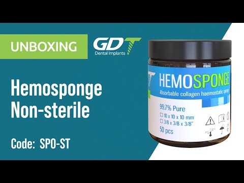
{"description":"This reveal showcases Hemosponge Non‑Sterile, a collagen sponge for general surgical applications. A recycled cardboard box opens to a sealed pouch containing a 50 × 50 × 10 mm sheet. ","media_image":"https:\/\/img.youtube.com\/vi\/KJGkwhixDvI\/hqdefault.jpg","media_link":"https:\/\/www.youtube.com\/watch?v=KJGkwhixDvI","media_page_content":{"type":"root","children":[{"type":"paragraph","children":[{"type":"text","value":"This reveal showcases Hemosponge Non‑Sterile, a collagen sponge for general surgical applications. A recycled cardboard box opens to a sealed pouch containing a 50 × 50 × 10 mm sheet. Absorption testing shows it can soak 28 times its weight. The host notes it is intended for trimming to size in large surgical fields and bulk hospital supply. A periodontal cross‑incision demo shows gentle compression stopping bleeding within 40 seconds; removal after three minutes leaves no adhesion. Cost‑flow slides cite a 45 % savings over single‑use sterile collagen sponges. The finale stresses that sterilization is required before use, compatible with autoclave or ETO gas methods."}]}]},"title":"Unboxing GDT Hemosponge - Absorbable Non-Sterile Sponge"}
Unboxing GDT Hemosponge - Absorbable Non-Sterile Sponge
This reveal showcases Hemosponge Non‑Sterile, a collagen sponge for general surgical applications. A recycled cardboard box opens to a sealed pouch containing a 50 × 50 × 10 mm sheet.
{"description":"This unboxing features GDT’s GBR Bone Tack Screw. The arrow‑shaped tip is calcium‑phosphate‑coated to promote osseointegration after removal. ","media_image":"https:\/\/i.ytimg.com\/vi\/Bqs-d1i2gTo\/hqdefault.jpg","media_link":"https:\/\/www.youtube.com\/watch?v=Bqs-d1i2gTo","media_page_content":{"type":"root","children":[{"type":"paragraph","children":[{"type":"text","value":"This unboxing features GDT’s GBR Bone Tack Screw. The arrow‑shaped tip is calcium‑phosphate‑coated to promote osseointegration after removal. The click‑in system includes a magnetic holder for one‑handed insertion. A synthetic cortical pull‑out test registers 68 N resistance. The video covers a lateral sinus window where the screw anchors a BioMembrane edge with a single tap. An in‑vitro µCT scan reveals trabecular islands forming around CaP particles. The package contains ten screws, with a rigid poly‑tray preventing needle deformation. A closing QR code links to a step‑by‑step video guide."}]}]},"title":"Unboxing GDT GBR Bone Tack Screw"}

Unboxing GDT GBR Bone Tack Screw
This unboxing features GDT’s GBR Bone Tack Screw. The arrow‑shaped tip is calcium‑phosphate‑coated to promote osseointegration after removal.
{"description":"The video opens a PGA Braided 5‑0 pack in violet. A 3\/8 c needle with silk‑glide coating shows smooth penetration. Tensile testing registers 0.9 kgf, meeting USP specs. A slide notes 50 % absorption at 14 days and full resorption at 90 days. A clinical clip places interrupted horizontal mattress sutures at a GBR site; patient feedback indicates high comfort. Packaging includes a barcode sticker for record entry. A clinician notes minimal inflammatory response compared with chromic gut.","media_image":"https:\/\/i.ytimg.com\/vi\/EUBWahyC8H0\/hq720.jpg","media_link":"https:\/\/www.youtube.com\/watch?v=EUBWahyC8H0","media_page_content":{"type":"root","children":[{"type":"paragraph","children":[{"type":"text","value":"The video opens a PGA Braided 5‑0 pack in violet. A 3\/8 c needle with silk‑glide coating shows smooth penetration. Tensile testing registers 0.9 kgf, meeting USP specs. A slide notes 50 % absorption at 14 days and full resorption at 90 days. A clinical clip places interrupted horizontal mattress sutures at a GBR site; patient feedback indicates high comfort. Packaging includes a barcode sticker for record entry. A clinician notes minimal inflammatory response compared with chromic gut."}]}]},"title":"Unboxing GDT PGA Suture"}

Unboxing GDT PGA Suture
The video opens a PGA Braided 5‑0 pack in violet. A 3/8 c needle with silk‑glide coating shows smooth penetration. Tensile testing registers 0.9 kgf, meeting USP specs. A slide notes 50 % absorption at 14 days and full resorption at 90 days. A clinical clip places interrupted horizontal mattress sutures at a GBR site; patient feedback indicates high comfort. Packaging includes a barcode sticker for record entry. A clinician notes minimal inflammatory response compared with chromic gut.
{"description":"The video opens a Nylon Monofilament 6‑0 box. A 3\/8 c needle with TEFLON coating reduces insertion force by 22 %. Tensile testing shows 1.25 kgf strength. A surgical host performs a simple‑interrupted stitch on a crestal incision, demonstrating zero memory during tie‑down. A slide notes zero absorption—suture removal on day 7. A comparison clip shows less inflammation than silk. Packaging includes a plastic cutter and a patient removal‑date pad. FDA 510(k) clearance appears in an overlay.","media_image":"https:\/\/i.ytimg.com\/vi\/K0Oe2_TX65U\/hq720.jpg","media_link":"https:\/\/www.youtube.com\/embed\/K0Oe2_TX65U","media_page_content":{"type":"root","children":[{"type":"paragraph","children":[{"type":"text","value":"The video opens a Nylon Monofilament 6‑0 box. A 3\/8 c needle with TEFLON coating reduces insertion force by 22 %. Tensile testing shows 1.25 kgf strength. A surgical host performs a simple‑interrupted stitch on a crestal incision, demonstrating zero memory during tie‑down. A slide notes zero absorption—suture removal on day 7. A comparison clip shows less inflammation than silk. Packaging includes a plastic cutter and a patient removal‑date pad. FDA 510(k) clearance appears in an overlay."}]}]},"title":"Unboxing GDT Nylon Suture"}

Unboxing GDT Nylon Suture
The video opens a Nylon Monofilament 6‑0 box. A 3/8 c needle with TEFLON coating reduces insertion force by 22 %. Tensile testing shows 1.25 kgf strength. A surgical host performs a simple‑interrupted stitch on a crestal incision, demonstrating zero memory during tie‑down. A slide notes zero absorption—suture removal on day 7. A comparison clip shows less inflammation than silk. Packaging includes a plastic cutter and a patient removal‑date pad. FDA 510(k) clearance appears in an overlay.
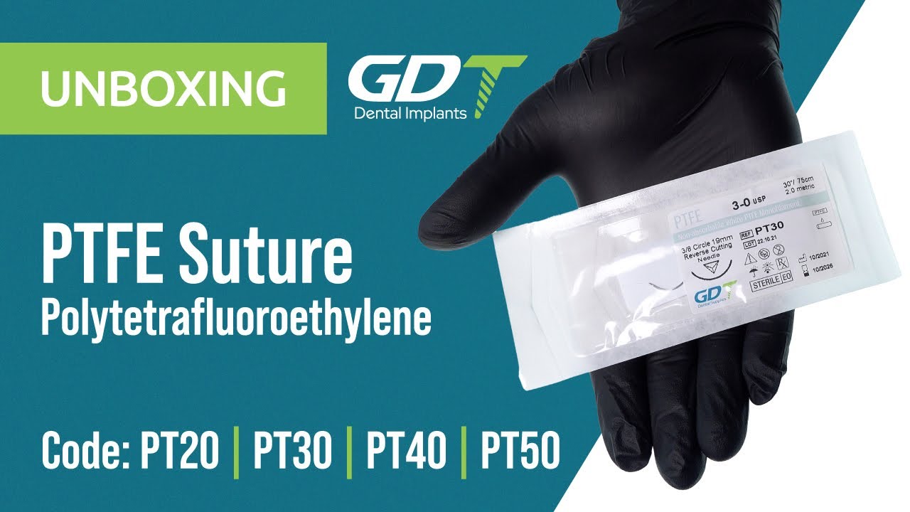
{"description":"The video opens a silver‑white PTFE Monofilament 6‑0 box. A 16 mm 3\/8 c TiN‑coated needle ensures radiographic visibility. Drag‑force tests show a 25 % reduction versus nylon. The surgical host notes the non‑absorbable suture is removed on day 10 but displays minimal bacterial wicking. SEM imagery reveals ultra‑smooth surfaces. A clinical segment shows a perio sling securing gingival margins. Higher cost is offset by reduced need for post‑op antibiotics. Packaging includes an integrated cutter and FDA Class II clearance. An automatic SMS reminder helps clinics schedule removal.","media_image":"https:\/\/img.youtube.com\/vi\/fv0s6_XjWLc\/maxresdefault.jpg","media_link":"https:\/\/www.youtube.com\/watch?v=fv0s6_XjWLc","media_page_content":{"type":"root","children":[{"type":"paragraph","children":[{"type":"text","value":"The video opens a silver‑white PTFE Monofilament 6‑0 box. A 16 mm 3\/8 c TiN‑coated needle ensures radiographic visibility. Drag‑force tests show a 25 % reduction versus nylon. The surgical host notes the non‑absorbable suture is removed on day 10 but displays minimal bacterial wicking. SEM imagery reveals ultra‑smooth surfaces. A clinical segment shows a perio sling securing gingival margins. Higher cost is offset by reduced need for post‑op antibiotics. Packaging includes an integrated cutter and FDA Class II clearance. An automatic SMS reminder helps clinics schedule removal."}]}]},"title":"Unboxing GDT PTFE Suture"}
Unboxing GDT PTFE Suture
The video opens a silver‑white PTFE Monofilament 6‑0 box. A 16 mm 3/8 c TiN‑coated needle ensures radiographic visibility. Drag‑force tests show a 25 % reduction versus nylon. The surgical host notes the non‑absorbable suture is removed on day 10 but displays minimal bacterial wicking. SEM imagery reveals ultra‑smooth surfaces. A clinical segment shows a perio sling securing gingival margins. Higher cost is offset by reduced need for post‑op antibiotics. Packaging includes an integrated cutter and FDA Class II clearance. An automatic SMS reminder helps clinics schedule removal.
{"description":"The video features PLX Monofilament 5‑0, an enhanced polydioxanone suture with e‑PTFE filler for added flexibility. A clear PET box reveals a pink color band for instant ID. Elongation testing shows 18 % stretch before failure, providing compliance in high‑tension sites. A lab host demonstrates a figure‑eight stitch during GBR, noting in‑vivo studies showing 40 % tensile loss at day 21 and full resorption by 180 days. A pH‑variation chart indicates stability between 6.3–6.8, reducing inflammation. A Paris surgeon reports lower patient discomfort compared with braided PGA. The clip ends with a QR code linking to integrated sterilization guidelines and a modular OR storage tray.","media_image":"https:\/\/img.youtube.com\/vi\/T7SlupYNjeI\/maxresdefault.jpg","media_link":"https:\/\/www.youtube.com\/watch?v=T7SlupYNjeI","media_page_content":{"type":"root","children":[{"type":"paragraph","children":[{"type":"text","value":"The video features PLX Monofilament 5‑0, an enhanced polydioxanone suture with e‑PTFE filler for added flexibility. A clear PET box reveals a pink color band for instant ID. Elongation testing shows 18 % stretch before failure, providing compliance in high‑tension sites. A lab host demonstrates a figure‑eight stitch during GBR, noting in‑vivo studies showing 40 % tensile loss at day 21 and full resorption by 180 days. A pH‑variation chart indicates stability between 6.3–6.8, reducing inflammation. A Paris surgeon reports lower patient discomfort compared with braided PGA. The clip ends with a QR code linking to integrated sterilization guidelines and a modular OR storage tray."}]}]},"title":"Unboxing GDT PLX Suture"}

Unboxing GDT PLX Suture
The video features PLX Monofilament 5‑0, an enhanced polydioxanone suture with e‑PTFE filler for added flexibility. A clear PET box reveals a pink color band for instant ID. Elongation testing shows 18 % stretch before failure, providing compliance in high‑tension sites. A lab host demonstrates a figure‑eight stitch during GBR, noting in‑vivo studies showing 40 % tensile loss at day 21 and full resorption by 180 days. A pH‑variation chart indicates stability between 6.3–6.8, reducing inflammation. A Paris surgeon reports lower patient discomfort compared with braided PGA. The clip ends with a QR code linking to integrated sterilization guidelines and a modular OR storage tray.
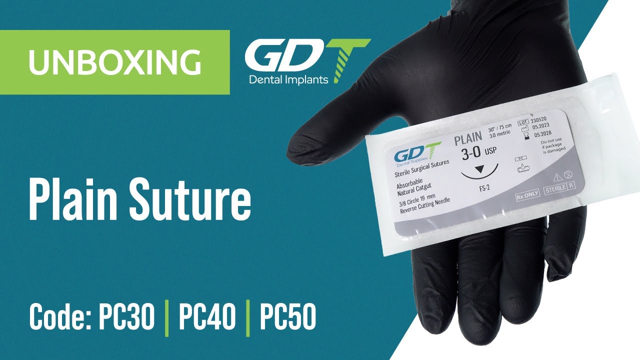
{"description":"The video unboxes Plain Gut 4‑0, a naturally absorbable surgical suture. A green pack opens to twin sterile pouches; each 45 cm strand is attached to a 3\/8 c 300S stainless‑steel needle with a silicon‑glide coating. Instron tensile testing shows an average 0.8 kgf breaking load—20 % above USP requirements. The surgical host demonstrates a simple interrupted stitch in the lower jaw: partial absorption occurs by day 7, complete resorption by week 21, with negligible inflammatory response. An animation details collagen breakdown by collagenases and macrophages. A pediatric dentist from California notes convenience in young patients since no removal is required. A cost slide indicates 30 % savings over synthetic absorbable sutures. The video concludes with CE and FDA 510(k) compliance, and the box contains a documentation card for digital chart integration.","media_image":"https:\/\/img.youtube.com\/vi\/DsYeyWscuOM\/maxresdefault.jpg","media_link":"https:\/\/www.youtube.com\/watch?v=DsYeyWscuOM","media_page_content":{"type":"root","children":[{"type":"paragraph","children":[{"type":"text","value":"The video unboxes Plain Gut 4‑0, a naturally absorbable surgical suture. A green pack opens to twin sterile pouches; each 45 cm strand is attached to a 3\/8 c 300S stainless‑steel needle with a silicon‑glide coating. Instron tensile testing shows an average 0.8 kgf breaking load—20 % above USP requirements. The surgical host demonstrates a simple interrupted stitch in the lower jaw: partial absorption occurs by day 7, complete resorption by week 21, with negligible inflammatory response. An animation details collagen breakdown by collagenases and macrophages. A pediatric dentist from California notes convenience in young patients since no removal is required. A cost slide indicates 30 % savings over synthetic absorbable sutures. The video concludes with CE and FDA 510(k) compliance, and the box contains a documentation card for digital chart integration."}]}]},"title":"Unboxing GDT Plain Suture"}
Unboxing GDT Plain Suture
The video unboxes Plain Gut 4‑0, a naturally absorbable surgical suture. A green pack opens to twin sterile pouches; each 45 cm strand is attached to a 3/8 c 300S stainless‑steel needle with a silicon‑glide coating. Instron tensile testing shows an average 0.8 kgf breaking load—20 % above USP requirements. The surgical host demonstrates a simple interrupted stitch in the lower jaw: partial absorption occurs by day 7, complete resorption by week 21, with negligible inflammatory response. An animation details collagen breakdown by collagenases and macrophages. A pediatric dentist from California notes convenience in young patients since no removal is required. A cost slide indicates 30 % savings over synthetic absorbable sutures. The video concludes with CE and FDA 510(k) compliance, and the box contains a documentation card for digital chart integration.
{"description":"Opening Rapid PGA 4‑0, a synthetic braided suture that loses half its strength by day 10 and resorbs in 42 days. Knot‑security testing records 90 % in two throws. Slow‑motion captures a surgeon’s knot without fraying. An Oslo clinician says uniform diameter simplifies suturing delicate peri‑oral tissues. Histology shows mild inflammation. A cost slide notes 15 % savings versus Vicryl Rapide. A yellow color code aids tray sorting. The video ends noting compatibility with interchangeable needle assortments.","media_image":"https:\/\/i.ytimg.com\/vi\/Dh8CGSZhbZc\/hq720.jpg","media_link":"https:\/\/www.youtube.com\/watch?v=Dh8CGSZhbZc","media_page_content":{"type":"root","children":[{"type":"paragraph","children":[{"type":"text","value":"Opening Rapid PGA 4‑0, a synthetic braided suture that loses half its strength by day 10 and resorbs in 42 days. Knot‑security testing records 90 % in two throws. Slow‑motion captures a surgeon’s knot without fraying. An Oslo clinician says uniform diameter simplifies suturing delicate peri‑oral tissues. Histology shows mild inflammation. A cost slide notes 15 % savings versus Vicryl Rapide. A yellow color code aids tray sorting. The video ends noting compatibility with interchangeable needle assortments."}]}]},"title":"Unboxing GDT RPGA Suture"}

Unboxing GDT RPGA Suture
Opening Rapid PGA 4‑0, a synthetic braided suture that loses half its strength by day 10 and resorbs in 42 days. Knot‑security testing records 90 % in two throws. Slow‑motion captures a surgeon’s knot without fraying. An Oslo clinician says uniform diameter simplifies suturing delicate peri‑oral tissues. Histology shows mild inflammation. A cost slide notes 15 % savings versus Vicryl Rapide. A yellow color code aids tray sorting. The video ends noting compatibility with interchangeable needle assortments.
{"description":"The video spotlights Silk Braided 5‑0, a classic non‑absorbable suture. A PE pouch opens to a 19 mm 3\/8 c needle. The host notes wax coating reduces capillarity, and polished black silk increases braid density. Quantitative inflammation shows moderate response at day 7, subsiding by day 14. In a posterior flap closure, the suture demonstrates strong knot retention. Low cost makes it a teaching‑hospital staple. A safety slide reminds removal on day 10. Packaging includes a QR code linking to Russian and Spanish tutorial videos.","media_image":"https:\/\/i.ytimg.com\/vi\/qAIg_Q3LDnk\/hq720.jpg","media_link":"https:\/\/www.youtube.com\/watch?v=qAIg_Q3LDnk","media_page_content":{"type":"root","children":[{"type":"paragraph","children":[{"type":"text","value":"The video spotlights Silk Braided 5‑0, a classic non‑absorbable suture. A PE pouch opens to a 19 mm 3\/8 c needle. The host notes wax coating reduces capillarity, and polished black silk increases braid density. Quantitative inflammation shows moderate response at day 7, subsiding by day 14. In a posterior flap closure, the suture demonstrates strong knot retention. Low cost makes it a teaching‑hospital staple. A safety slide reminds removal on day 10. Packaging includes a QR code linking to Russian and Spanish tutorial videos."}]}]},"title":"Unboxing GDT Silk Suture"}

Unboxing GDT Silk Suture
The video spotlights Silk Braided 5‑0, a classic non‑absorbable suture. A PE pouch opens to a 19 mm 3/8 c needle. The host notes wax coating reduces capillarity, and polished black silk increases braid density. Quantitative inflammation shows moderate response at day 7, subsiding by day 14. In a posterior flap closure, the suture demonstrates strong knot retention. Low cost makes it a teaching‑hospital staple. A safety slide reminds removal on day 10. Packaging includes a QR code linking to Russian and Spanish tutorial videos.
{"description":"The video features Bovine Sponge Plug, an 8 × 10 mm screw‑shaped bovine collagen. Compression testing shows 20 % collapse at 100 N. An animation highlights a diagonal matrix creating blood reservoirs. A Tirana surgeon fills an extraction socket and secures with a cross suture. Three‑month CBCT reveals ridge‑height preservation. Cost slides show 25 % savings over synthetic plugs. The sterile double pack carries a 3‑year shelf life.","media_image":"https:\/\/i.ytimg.com\/vi\/fMbOxFCchmU\/hq720.jpg","media_link":"https:\/\/www.youtube.com\/watch?v=fMbOxFCchmU","media_page_content":{"type":"root","children":[{"type":"paragraph","children":[{"type":"text","value":"The video features Bovine Sponge Plug, an 8 × 10 mm screw‑shaped bovine collagen. Compression testing shows 20 % collapse at 100 N. An animation highlights a diagonal matrix creating blood reservoirs. A Tirana surgeon fills an extraction socket and secures with a cross suture. Three‑month CBCT reveals ridge‑height preservation. Cost slides show 25 % savings over synthetic plugs. The sterile double pack carries a 3‑year shelf life."}]}]},"title":"Unboxing GDT Sponge Bovine Grafting Plug"}

Unboxing GDT Sponge Bovine Grafting Plug
The video features Bovine Sponge Plug, an 8 × 10 mm screw‑shaped bovine collagen. Compression testing shows 20 % collapse at 100 N. An animation highlights a diagonal matrix creating blood reservoirs. A Tirana surgeon fills an extraction socket and secures with a cross suture. Three‑month CBCT reveals ridge‑height preservation. Cost slides show 25 % savings over synthetic plugs. The sterile double pack carries a 3‑year shelf life.
{"description":"A synthetic β‑TCP\/collagen plug, 70\/30 ratio, 7 mm diameter. In‑vitro testing shows 26‑fold blood absorption. The host highlights accelerated calcium‑ion release. A maxillary‑anterior clip inserts the plug with a single suture. Osteoblast density increases 40 % at 14 days. Mid‑range pricing; eco‑friendly wrap completes the package.","media_image":"https:\/\/i.ytimg.com\/vi\/E2Bz6ZRyXLM\/hq720.jpg","media_link":"https:\/\/www.youtube.com\/watch?v=E2Bz6ZRyXLM","media_page_content":{"type":"root","children":[{"type":"paragraph","children":[{"type":"text","value":"A synthetic β‑TCP\/collagen plug, 70\/30 ratio, 7 mm diameter. In‑vitro testing shows 26‑fold blood absorption. The host highlights accelerated calcium‑ion release. A maxillary‑anterior clip inserts the plug with a single suture. Osteoblast density increases 40 % at 14 days. Mid‑range pricing; eco‑friendly wrap completes the package."}]}]},"title":"Unboxing GDT Sponge Grafting Plug"}

Unboxing GDT Sponge Grafting Plug
A synthetic β‑TCP/collagen plug, 70/30 ratio, 7 mm diameter. In‑vitro testing shows 26‑fold blood absorption. The host highlights accelerated calcium‑ion release. A maxillary‑anterior clip inserts the plug with a single suture. Osteoblast density increases 40 % at 14 days. Mid‑range pricing; eco‑friendly wrap completes the package.
{"description":"The box holds two 5 × 10 mm β‑TCP cones. The host shows 120 % volume expansion on moisture contact. An animation demonstrates press‑fit for crestal sinus lift. A Rome specialist presents 6‑month CBCT with full cortical fusion. A slide notes time saved over autogenous graft harvesting. CE‑marked and MRI‑compatible.","media_image":"https:\/\/i.ytimg.com\/vi\/GAWi3sk-eLQ\/hq720.jpg","media_link":"https:\/\/www.youtube.com\/watch?v=GAWi3sk-eLQ","media_page_content":{"type":"root","children":[{"type":"paragraph","children":[{"type":"text","value":"The box holds two 5 × 10 mm β‑TCP cones. The host shows 120 % volume expansion on moisture contact. An animation demonstrates press‑fit for crestal sinus lift. A Rome specialist presents 6‑month CBCT with full cortical fusion. A slide notes time saved over autogenous graft harvesting. CE‑marked and MRI‑compatible."}]}]},"title":"Unboxing GDT Synthetic Bone Graft Cones - 2 Pieces"}

Unboxing GDT Synthetic Bone Graft Cones - 2 Pieces
The box holds two 5 × 10 mm β‑TCP cones. The host shows 120 % volume expansion on moisture contact. An animation demonstrates press‑fit for crestal sinus lift. A Rome specialist presents 6‑month CBCT with full cortical fusion. A slide notes time saved over autogenous graft harvesting. CE‑marked and MRI‑compatible.
{"description":"The presenter opens a compact cardboard box and lifts out two precision‑milled 10 × 10 × 10 mm cubes of β‑TCP and hydroxyapatite. A close‑up reveals a marble‑like porosity—an intricate network of micro‑channels that multiplies surface area and speeds vascular ingrowth. A quick scale demo shows each cube soaking up saline and gaining 50 % weight within an hour, proof of its protein‑wicking capacity. Next, the host trims a cube with a sterile blade to illustrate how easily it can be customised for narrow ridges or irregular defects. A six‑month CBCT from an Italian surgeon displays flawless osseous integration: the graft‑to‑bone interface has completely disappeared. Practical tip: pre‑soak in saline for a snug press‑fit. The package bears a CE mark and a three‑year shelf life, plus a QR code linking to a full surgical guide. A closing slide quantifies benefits—shorter operating time and zero donor‑site morbidity—translating into less pain for the patient and lower overall cost.","media_image":"https:\/\/i.ytimg.com\/vi\/LoX0OmFbN7s\/hq720.jpg","media_link":"https:\/\/www.youtube.com\/watch?v=LoX0OmFbN7s","media_page_content":{"type":"root","children":[{"type":"paragraph","children":[{"type":"text","value":"The presenter opens a compact cardboard box and lifts out two precision‑milled 10 × 10 × 10 mm cubes of β‑TCP and hydroxyapatite. A close‑up reveals a marble‑like porosity—an intricate network of micro‑channels that multiplies surface area and speeds vascular ingrowth. A quick scale demo shows each cube soaking up saline and gaining 50 % weight within an hour, proof of its protein‑wicking capacity. Next, the host trims a cube with a sterile blade to illustrate how easily it can be customised for narrow ridges or irregular defects. A six‑month CBCT from an Italian surgeon displays flawless osseous integration: the graft‑to‑bone interface has completely disappeared. Practical tip: pre‑soak in saline for a snug press‑fit. The package bears a CE mark and a three‑year shelf life, plus a QR code linking to a full surgical guide. A closing slide quantifies benefits—shorter operating time and zero donor‑site morbidity—translating into less pain for the patient and lower overall cost."}]}]},"title":"Unboxing GDT Synthetic Bone Graft Cubes - 2 Pieces"}

Unboxing GDT Synthetic Bone Graft Cubes - 2 Pieces
The presenter opens a compact cardboard box and lifts out two precision‑milled 10 × 10 × 10 mm cubes of β‑TCP and hydroxyapatite. A close‑up reveals a marble‑like porosity—an intricate network of micro‑channels that multiplies surface area and speeds vascular ingrowth. A quick scale demo shows each cube soaking up saline and gaining 50 % weight within an hour, proof of its protein‑wicking capacity. Next, the host trims a cube with a sterile blade to illustrate how easily it can be customised for narrow ridges or irregular defects. A six‑month CBCT from an Italian surgeon displays flawless osseous integration: the graft‑to‑bone interface has completely disappeared. Practical tip: pre‑soak in saline for a snug press‑fit. The package bears a CE mark and a three‑year shelf life, plus a QR code linking to a full surgical guide. A closing slide quantifies benefits—shorter operating time and zero donor‑site morbidity—translating into less pain for the patient and lower overall cost.
{"description":"Removing the clear lid reveals four 6 × 6 × 6 mm β‑TCP cubes that clink onto a stainless tray, their hollow sound hinting at high porosity. A digital press applies force, confirming 7 MPa compressive strength—enough to handle early functional load without collapse. A 3‑D animation shows the cubes stacking like LEGO bricks, rapidly rebuilding a ridge lost to trauma. A clinical clip features a Slovenian surgeon performing a two‑stage ridge split: cubes are inserted into the gap, covered with a GBR membrane, and within three months the ridge unifies with minimal mineral resorption. Key advantage: surgical flexibility—use exactly the number of cubes you need, waste less material, and adapt to complex shapes on the fly. The rigid tray can be re‑sterilised, and an MRI‑Safe label guarantees artifact‑free imaging post‑op. A QR code links to a tutorial on advanced placement tactics. The closing cost slide shows a 20 % saving versus a single large block, both in material expenditures and shaping time.","media_image":"https:\/\/i.ytimg.com\/vi\/4FvTGCw-iGw\/hq720.jpg","media_link":"https:\/\/www.youtube.com\/watch?v=4FvTGCw-iGw","media_page_content":{"type":"root","children":[{"type":"paragraph","children":[{"type":"text","value":"Removing the clear lid reveals four 6 × 6 × 6 mm β‑TCP cubes that clink onto a stainless tray, their hollow sound hinting at high porosity. A digital press applies force, confirming 7 MPa compressive strength—enough to handle early functional load without collapse. A 3‑D animation shows the cubes stacking like LEGO bricks, rapidly rebuilding a ridge lost to trauma. A clinical clip features a Slovenian surgeon performing a two‑stage ridge split: cubes are inserted into the gap, covered with a GBR membrane, and within three months the ridge unifies with minimal mineral resorption. Key advantage: surgical flexibility—use exactly the number of cubes you need, waste less material, and adapt to complex shapes on the fly. The rigid tray can be re‑sterilised, and an MRI‑Safe label guarantees artifact‑free imaging post‑op. A QR code links to a tutorial on advanced placement tactics. The closing cost slide shows a 20 % saving versus a single large block, both in material expenditures and shaping time."}]}]},"title":"Unboxing GDT Synthetic Bone Graft Cubes - 4 Pieces"}

Unboxing GDT Synthetic Bone Graft Cubes - 4 Pieces
Removing the clear lid reveals four 6 × 6 × 6 mm β‑TCP cubes that clink onto a stainless tray, their hollow sound hinting at high porosity. A digital press applies force, confirming 7 MPa compressive strength—enough to handle early functional load without collapse. A 3‑D animation shows the cubes stacking like LEGO bricks, rapidly rebuilding a ridge lost to trauma. A clinical clip features a Slovenian surgeon performing a two‑stage ridge split: cubes are inserted into the gap, covered with a GBR membrane, and within three months the ridge unifies with minimal mineral resorption. Key advantage: surgical flexibility—use exactly the number of cubes you need, waste less material, and adapt to complex shapes on the fly. The rigid tray can be re‑sterilised, and an MRI‑Safe label guarantees artifact‑free imaging post‑op. A QR code links to a tutorial on advanced placement tactics. The closing cost slide shows a 20 % saving versus a single large block, both in material expenditures and shaping time.
{"description":"A shiny foil pouch snaps open, releasing a stream of 0.5–1 mm β‑TCP granules into an ice‑blue Petri dish. Macro photography reveals tiny octagonal crystals sparkling under cool light. The lab host cites a specific surface area of 85 m² per gram—a playground for osteoblasts. A clear Luer‑Lock syringe demonstrates gentle injection into a mandibular three‑wall defect, avoiding air pockets. A collagen plug is tamped over to secure volume. Four‑month CBCT shows uniform mineral density, free of voids or radiolucencies. Packaging is fully compostable within six months, aligning with GDT’s Green Dental policy. A closing chart highlights a 30 % savings in material cost and reduced OR waste compared with pure HA granules. The CE mark is prominent, and the QR code links to the safety data sheet and protocol pages.","media_image":"https:\/\/i.ytimg.com\/vi\/wv7qYxJWWP0\/hq720.jpg","media_link":"https:\/\/www.youtube.com\/watch?v=wv7qYxJWWP0","media_page_content":{"type":"root","children":[{"type":"paragraph","children":[{"type":"text","value":"A shiny foil pouch snaps open, releasing a stream of 0.5–1 mm β‑TCP granules into an ice‑blue Petri dish. Macro photography reveals tiny octagonal crystals sparkling under cool light. The lab host cites a specific surface area of 85 m² per gram—a playground for osteoblasts. A clear Luer‑Lock syringe demonstrates gentle injection into a mandibular three‑wall defect, avoiding air pockets. A collagen plug is tamped over to secure volume. Four‑month CBCT shows uniform mineral density, free of voids or radiolucencies. Packaging is fully compostable within six months, aligning with GDT’s Green Dental policy. A closing chart highlights a 30 % savings in material cost and reduced OR waste compared with pure HA granules. The CE mark is prominent, and the QR code links to the safety data sheet and protocol pages."}]}]},"title":"Unboxing GDT Synthetic Bone Graft Granules"}

Unboxing GDT Synthetic Bone Graft Granules
A shiny foil pouch snaps open, releasing a stream of 0.5–1 mm β‑TCP granules into an ice‑blue Petri dish. Macro photography reveals tiny octagonal crystals sparkling under cool light. The lab host cites a specific surface area of 85 m² per gram—a playground for osteoblasts. A clear Luer‑Lock syringe demonstrates gentle injection into a mandibular three‑wall defect, avoiding air pockets. A collagen plug is tamped over to secure volume. Four‑month CBCT shows uniform mineral density, free of voids or radiolucencies. Packaging is fully compostable within six months, aligning with GDT’s Green Dental policy. A closing chart highlights a 30 % savings in material cost and reduced OR waste compared with pure HA granules. The CE mark is prominent, and the QR code links to the safety data sheet and protocol pages.
{"description":"A transparent single‑use syringe holds 1.5 cc of β‑TCP‑collagen granules, sealed in a rigid blister. The dual‑stage plunger creates a gentle vacuum, preventing air pockets. A tantalum X‑ray marker embedded in the barrel clearly indicates graft location and volume on post‑op radiographs. In a Madrid clinic, a surgeon injects the granules to augment a mandibular onlay, using slow, controlled pressure for even dispersion. Immediate intra‑oral scanning maps the site, confirming a void‑free fill. A finance slide notes a 15‑minute reduction in OR time, lower staff costs, and zero cross‑contamination risks thanks to the disposable system. Shelf life is three years; packaging is recyclable and printed with plant‑based ink. A QR code links to GDT’s advanced injection webinar, covering membrane pairing tips and plunger torque metrics.","media_image":"https:\/\/i.ytimg.com\/vi\/tIbFCZAnRxg\/hq720.jpg","media_link":"https:\/\/www.youtube.com\/watch?v=tIbFCZAnRxg","media_page_content":{"type":"root","children":[{"type":"paragraph","children":[{"type":"text","value":"A transparent single‑use syringe holds 1.5 cc of β‑TCP‑collagen granules, sealed in a rigid blister. The dual‑stage plunger creates a gentle vacuum, preventing air pockets. A tantalum X‑ray marker embedded in the barrel clearly indicates graft location and volume on post‑op radiographs. In a Madrid clinic, a surgeon injects the granules to augment a mandibular onlay, using slow, controlled pressure for even dispersion. Immediate intra‑oral scanning maps the site, confirming a void‑free fill. A finance slide notes a 15‑minute reduction in OR time, lower staff costs, and zero cross‑contamination risks thanks to the disposable system. Shelf life is three years; packaging is recyclable and printed with plant‑based ink. A QR code links to GDT’s advanced injection webinar, covering membrane pairing tips and plunger torque metrics."}]}]},"title":"Unboxing GDT Synthetic Bone Graft Syringe"}

Unboxing GDT Synthetic Bone Graft Syringe
A transparent single‑use syringe holds 1.5 cc of β‑TCP‑collagen granules, sealed in a rigid blister. The dual‑stage plunger creates a gentle vacuum, preventing air pockets. A tantalum X‑ray marker embedded in the barrel clearly indicates graft location and volume on post‑op radiographs. In a Madrid clinic, a surgeon injects the granules to augment a mandibular onlay, using slow, controlled pressure for even dispersion. Immediate intra‑oral scanning maps the site, confirming a void‑free fill. A finance slide notes a 15‑minute reduction in OR time, lower staff costs, and zero cross‑contamination risks thanks to the disposable system. Shelf life is three years; packaging is recyclable and printed with plant‑based ink. A QR code links to GDT’s advanced injection webinar, covering membrane pairing tips and plunger torque metrics.
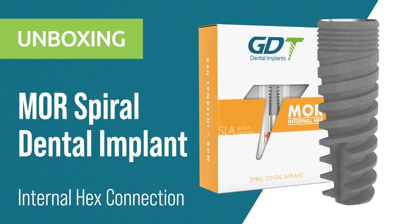
{"description":"Join us for an in-depth unboxing of GDT MOR Spiral Dental Implant. Featuring a tapered body with aggressive apical threads and self-drilling capabilities, the MOR Implant is optimized for a wide range of clinical indications, including immediate loading.\n\nManufactured from Titanium Grade 5 (Ti-6Al-4V Eli) with SLA (Sandblast, Large Grit, Acid-etch) surface treatment for improved osseointegration, this implant delivers excellent primary stability and long-term success.","media_image":"https:\/\/i.ytimg.com\/vi\/km8E5IRYXiA\/hq720.jpg","media_link":"https:\/\/www.youtube.com\/watch?v=km8E5IRYXiA","media_page_content":{"type":"root","children":[{"type":"paragraph","children":[{"type":"text","value":"Join us for an in-depth unboxing of GDT MOR Spiral Dental Implant. Featuring a tapered body with aggressive apical threads and self-drilling capabilities, the MOR Implant is optimized for a wide range of clinical indications, including immediate loading.\n\nManufactured from Titanium Grade 5 (Ti-6Al-4V Eli) with SLA (Sandblast, Large Grit, Acid-etch) surface treatment for improved osseointegration, this implant delivers excellent primary stability and long-term success.\n\n"}]}]},"title":"Unboxing MOR Spiral Implant | GDT Dental Implants™"}
Unboxing MOR Spiral Implant | GDT Dental Implants™
Join us for an in-depth unboxing of GDT MOR Spiral Dental Implant. Featuring a tapered body with aggressive apical threads and self-drilling capabilities, the MOR Implant is optimized for a wide range of clinical indications, including immediate loading.
Manufactured from Titanium Grade 5 (Ti-6Al-4V Eli) with SLA (Sandblast, Large Grit, Acid-etch) surface treatment for improved osseointegration, this implant delivers excellent primary stability and long-term success.
{"description":"Join us for the unboxing of our ABA Spiral Dental Implant, engineered for versatility across all surgical protocols, including immediate loading, two-stage, and flapless procedures.\n\nGDT ABA Implant features a tapered body with deep, aggressive apical threads, optimized for engagement in narrow ridges and poor bone conditions, without the need for bone grafting.\n\nMade from Titanium Grade 5 (Ti-6Al-4V ELI) and treated with SLA (Sandblast, Large Grit, Acid-etch) for improved osseointegration, this implant supports excellent primary stability and effective load distribution.\n\nIdeal for dentists seeking efficient handling and high compatibility across ridge types and indications.","media_image":"https:\/\/i.ytimg.com\/vi\/TpKGuEWZudU\/hqdefault.jpg","media_link":"https:\/\/www.youtube.com\/watch?v=TpKGuEWZudU","media_page_content":{"type":"root","children":[{"type":"paragraph","children":[{"type":"text","value":"Join us for the unboxing of our ABA Spiral Dental Implant, engineered for versatility across all surgical protocols, including immediate loading, two-stage, and flapless procedures.\n\nGDT ABA Implant features a tapered body with deep, aggressive apical threads, optimized for engagement in narrow ridges and poor bone conditions, without the need for bone grafting.\n\nMade from Titanium Grade 5 (Ti-6Al-4V ELI) and treated with SLA (Sandblast, Large Grit, Acid-etch) for improved osseointegration, this implant supports excellent primary stability and effective load distribution.\n\nIdeal for dentists seeking efficient handling and high compatibility across ridge types and indications.\n"}]}]},"title":"Unboxing MOR Spiral Implant | GDT Dental Implants"}

Unboxing MOR Spiral Implant | GDT Dental Implants
Join us for the unboxing of our ABA Spiral Dental Implant, engineered for versatility across all surgical protocols, including immediate loading, two-stage, and flapless procedures.
GDT ABA Implant features a tapered body with deep, aggressive apical threads, optimized for engagement in narrow ridges and poor bone conditions, without the need for bone grafting.
Made from Titanium Grade 5 (Ti-6Al-4V ELI) and treated with SLA (Sandblast, Large Grit, Acid-etch) for improved osseointegration, this implant supports excellent primary stability and effective load distribution.
Ideal for dentists seeking efficient handling and high compatibility across ridge types and indications.
GDT Dental Implants
Get to know us
{"description":"This vibrant corporate film reveals GDT’s core philosophy: innovation, transparency, and global partnership. It intercuts close‑ups of clinician workshops in Rome, São Paulo, and Hong Kong with slow‑motion factory shots of titanium shavings dancing to an original symphonic score. Dual Hebrew‑English narration notes that the company reinvests 15 % of revenue in R\u0026D, covering Nano‑Etch surfaces and 10‑million‑cycle fatigue tests. Dentists praise the free G‑Academy virtual courses and subsidized hands‑on sessions. Humanitarian clips show GDT teams volunteering in Uganda, with patients beaming after fixed restorations. The final message—“The vision behind every smile is trust”—appears over a Tel Aviv sunset, inviting new partners to join the dental revolution.","media_image":"https:\/\/i.ytimg.com\/vi\/lDFx0wGQtaI\/hq720.jpg?sqp=-oaymwEnCNAFEJQDSFryq4qpAxkIARUAAIhCGAHYAQHiAQoIGBACGAY4AUAB\u0026rs=AOn4CLCQyA_TiGW2ZjHV-5-LnUDejUQ7pw","media_link":"https:\/\/www.youtube.com\/watch?v=lDFx0wGQtaI\u0026pp=ygUzVGhlIFZpc2lvbiBCZWhpbmQgRXZlcnkgU21pbGUgLSBHRFQgRGVudGFsIEltcGxhbnRz","media_page_content":{"type":"root","children":[{"type":"paragraph","children":[{"type":"text","value":"This vibrant corporate film reveals GDT’s core philosophy: innovation, transparency, and global partnership. It intercuts close‑ups of clinician workshops in Rome, São Paulo, and Hong Kong with slow‑motion factory shots of titanium shavings dancing to an original symphonic score. Dual Hebrew‑English narration notes that the company reinvests 15 % of revenue in R\u0026D, covering Nano‑Etch surfaces and 10‑million‑cycle fatigue tests. Dentists praise the free G‑Academy virtual courses and subsidized hands‑on sessions. Humanitarian clips show GDT teams volunteering in Uganda, with patients beaming after fixed restorations. The final message—“The vision behind every smile is trust”—appears over a Tel Aviv sunset, inviting new partners to join the dental revolution."}]}]},"title":" The Vision Behind Every Smile - GDT Dental Implants"}

The Vision Behind Every Smile - GDT Dental Implants
This vibrant corporate film reveals GDT’s core philosophy: innovation, transparency, and global partnership. It intercuts close‑ups of clinician workshops in Rome, São Paulo, and Hong Kong with slow‑motion factory shots of titanium shavings dancing to an original symphonic score. Dual Hebrew‑English narration notes that the company reinvests 15 % of revenue in R&D, covering Nano‑Etch surfaces and 10‑million‑cycle fatigue tests. Dentists praise the free G‑Academy virtual courses and subsidized hands‑on sessions. Humanitarian clips show GDT teams volunteering in Uganda, with patients beaming after fixed restorations. The final message—“The vision behind every smile is trust”—appears over a Tel Aviv sunset, inviting new partners to join the dental revolution.
{"description":"This video offers a rare, behind‑the‑scenes look at GDT Dental Implants’ manufacturing plant and headquarters. The tour opens in the company lobby, where GDT’s mission—making cutting‑edge implant solutions accessible worldwide—is introduced. The camera then moves to the R\u0026D suites, where engineers and clinicians stress‑test new implant geometries. Fully automated CNC lines mill medical‑grade titanium with microscopic precision; after ultrasonic cleaning, implants enter controlled‑atmosphere rooms to maintain sterility. The video highlights a double‑sterilization process and GDT’s distinctive Double Blister packaging, ensuring asepsis until chairside opening. Laser metrology and digital imaging verify full ISO and FDA compliance, while quality‑assurance staff emphasize a culture of innovation and continuous improvement. The tour ends in GDT’s training center, where dentists receive hands‑on instruction in advanced implant techniques—underscoring how shared knowledge drives better clinical outcomes and patient experiences.","media_image":"https:\/\/i.ytimg.com\/vi\/w_Nh4csMaTg\/hq720.jpg","media_link":"https:\/\/www.youtube.com\/watch?v=w_Nh4csMaTg","media_page_content":{"type":"root","children":[{"type":"paragraph","children":[{"type":"text","value":"This video offers a rare, behind‑the‑scenes look at GDT Dental Implants’ manufacturing plant and headquarters. The tour opens in the company lobby, where GDT’s mission—making cutting‑edge implant solutions accessible worldwide—is introduced. The camera then moves to the R\u0026D suites, where engineers and clinicians stress‑test new implant geometries. Fully automated CNC lines mill medical‑grade titanium with microscopic precision; after ultrasonic cleaning, implants enter controlled‑atmosphere rooms to maintain sterility. The video highlights a double‑sterilization process and GDT’s distinctive Double Blister packaging, ensuring asepsis until chairside opening. Laser metrology and digital imaging verify full ISO and FDA compliance, while quality‑assurance staff emphasize a culture of innovation and continuous improvement. The tour ends in GDT’s training center, where dentists receive hands‑on instruction in advanced implant techniques—underscoring how shared knowledge drives better clinical outcomes and patient experiences."}]}]},"title":"Behind-the-scenes Virtual Tour: GDT Dental Implants"}

Behind-the-scenes Virtual Tour: GDT Dental Implants
This video offers a rare, behind‑the‑scenes look at GDT Dental Implants’ manufacturing plant and headquarters. The tour opens in the company lobby, where GDT’s mission—making cutting‑edge implant solutions accessible worldwide—is introduced. The camera then moves to the R&D suites, where engineers and clinicians stress‑test new implant geometries. Fully automated CNC lines mill medical‑grade titanium with microscopic precision; after ultrasonic cleaning, implants enter controlled‑atmosphere rooms to maintain sterility. The video highlights a double‑sterilization process and GDT’s distinctive Double Blister packaging, ensuring asepsis until chairside opening. Laser metrology and digital imaging verify full ISO and FDA compliance, while quality‑assurance staff emphasize a culture of innovation and continuous improvement. The tour ends in GDT’s training center, where dentists receive hands‑on instruction in advanced implant techniques—underscoring how shared knowledge drives better clinical outcomes and patient experiences.
{"description":"This video chronicles a two‑day intensive workshop in Tirana where GDT specialists teach local dentists advanced prosthetic and restructuring protocols. Day 1 features a live demonstration of full‑arch reconstruction using angled Multi‑Unit Abutments and a split‑file CAD\/CAM technique. Close‑up footage shows an intra‑oral scan and a 3‑D‑printed surgical guide directing ridge reduction. Day 2 focuses on digital smile design: attendees plan full‑zirconia crowns mated to GDT Ti‑Bases. Guest lecturers from the U.S. and Germany highlight the RBM surface’s role in reducing marginal bone loss and present five‑year success rates above 98 %. Interviews with Albanian clinicians confirm that the comprehensive “surgery‑plus‑prosthetics” approach streamlines workflow and minimizes chairside adjustments. The workshop ends with a certificate ceremony, and participants learn about six months of free online mentoring that includes real‑case reviews and 48‑hour component delivery within the Albanian market.","media_image":"https:\/\/i.ytimg.com\/vi\/a0flpqJknLM\/hq720.jpg","media_link":"https:\/\/www.youtube.com\/watch?v=a0flpqJknLM\u0026pp=ygVNR0RUIERlbnRhbCBJbXBsYW50cyAtIFByb3N0aGV0aWNzICYgUmVzdHJ1Y3R1cmluZyBXb3Jrc2hvcCBhdCBUaXJhbmEsIEFsYmFuaWE%3D","media_page_content":{"type":"root","children":[{"type":"paragraph","children":[{"type":"text","value":"This video chronicles a two‑day intensive workshop in Tirana where GDT specialists teach local dentists advanced prosthetic and restructuring protocols. Day 1 features a live demonstration of full‑arch reconstruction using angled Multi‑Unit Abutments and a split‑file CAD\/CAM technique. Close‑up footage shows an intra‑oral scan and a 3‑D‑printed surgical guide directing ridge reduction. Day 2 focuses on digital smile design: attendees plan full‑zirconia crowns mated to GDT Ti‑Bases. Guest lecturers from the U.S. and Germany highlight the RBM surface’s role in reducing marginal bone loss and present five‑year success rates above 98 %. Interviews with Albanian clinicians confirm that the comprehensive “surgery‑plus‑prosthetics” approach streamlines workflow and minimizes chairside adjustments. The workshop ends with a certificate ceremony, and participants learn about six months of free online mentoring that includes real‑case reviews and 48‑hour component delivery within the Albanian market.\n"}]}]},"title":" GDT Dental Implants - Prosthetics \u0026 Restructuring Workshop at Tirana, Albania"}

GDT Dental Implants - Prosthetics & Restructuring Workshop at Tirana, Albania
This video chronicles a two‑day intensive workshop in Tirana where GDT specialists teach local dentists advanced prosthetic and restructuring protocols. Day 1 features a live demonstration of full‑arch reconstruction using angled Multi‑Unit Abutments and a split‑file CAD/CAM technique. Close‑up footage shows an intra‑oral scan and a 3‑D‑printed surgical guide directing ridge reduction. Day 2 focuses on digital smile design: attendees plan full‑zirconia crowns mated to GDT Ti‑Bases. Guest lecturers from the U.S. and Germany highlight the RBM surface’s role in reducing marginal bone loss and present five‑year success rates above 98 %. Interviews with Albanian clinicians confirm that the comprehensive “surgery‑plus‑prosthetics” approach streamlines workflow and minimizes chairside adjustments. The workshop ends with a certificate ceremony, and participants learn about six months of free online mentoring that includes real‑case reviews and 48‑hour component delivery within the Albanian market.
{"description":"This video introduces Martin Morra, GDT’s new regional sales manager for Spanish‑speaking markets. He outlines his 15‑year background in orthopedic fillers and dental restoration. Morra details plans to expand distributor networks across Latin America via Spanish‑language online trainings, portable demo kits, and logistic support from a Madrid hub. An animation summarizes the “3‑Tier Support” model—clinician, lab, patient—ensuring 24\/7 assistance. Morra highlights the availability of GDT’s Spanish training materials, including surgical guides and procedural sheets. He invites dentists to a webinar on the EVA implant and announces a new E‑shop that simplifies purchases in countries with import restrictions.","media_image":"https:\/\/i.ytimg.com\/vi\/zqkdcwGxTJY\/hqdefault.jpg","media_link":"https:\/\/www.youtube.com\/watch?v=zqkdcwGxTJY","media_page_content":{"type":"root","children":[{"type":"paragraph","children":[{"type":"text","value":"This video introduces Martin Morra, GDT’s new regional sales manager for Spanish‑speaking markets. He outlines his 15‑year background in orthopedic fillers and dental restoration. Morra details plans to expand distributor networks across Latin America via Spanish‑language online trainings, portable demo kits, and logistic support from a Madrid hub. An animation summarizes the “3‑Tier Support” model—clinician, lab, patient—ensuring 24\/7 assistance. Morra highlights the availability of GDT’s Spanish training materials, including surgical guides and procedural sheets. He invites dentists to a webinar on the EVA implant and announces a new E‑shop that simplifies purchases in countries with import restrictions."}]}]},"title":"Meet Martin Morra - Regional Sales Manager (Español) | GDT Dental Implants"}

Meet Martin Morra - Regional Sales Manager (Español) | GDT Dental Implants
This video introduces Martin Morra, GDT’s new regional sales manager for Spanish‑speaking markets. He outlines his 15‑year background in orthopedic fillers and dental restoration. Morra details plans to expand distributor networks across Latin America via Spanish‑language online trainings, portable demo kits, and logistic support from a Madrid hub. An animation summarizes the “3‑Tier Support” model—clinician, lab, patient—ensuring 24/7 assistance. Morra highlights the availability of GDT’s Spanish training materials, including surgical guides and procedural sheets. He invites dentists to a webinar on the EVA implant and announces a new E‑shop that simplifies purchases in countries with import restrictions.
{"description":"This video celebrates GDT’s recognition as Israel’s 2022 “Exporter of the Year.” It opens with an official ceremony attended by the Minister of Economy, highlighting the company’s 34 % year‑on‑year growth in overseas sales. Industrial footage shows CNC milling lines in Holon and a logistics hub in Ashdod processing over 15 000 monthly shipments to nearly 70 countries. The CEO outlines the strategy: competitive pricing through full vertical manufacturing, R\u0026D investment, and academic partnerships. Dentists from the U.S. and India praise rapid delivery times and multilingual technical support. Special focus is given to the G‑Care extended warranty, which offers a free replacement implant for any failure within five years. The video closes with employees hoisting the award trophy, declaring that “export drives innovation,” and pledging new breakthroughs for 2023.","media_image":"https:\/\/i.ytimg.com\/vi\/44iEaQ3ehWk\/hq720.jpg","media_link":"https:\/\/www.youtube.com\/watch?v=44iEaQ3ehWk","media_page_content":{"type":"root","children":[{"type":"paragraph","children":[{"type":"text","value":"This video celebrates GDT’s recognition as Israel’s 2022 “Exporter of the Year.” It opens with an official ceremony attended by the Minister of Economy, highlighting the company’s 34 % year‑on‑year growth in overseas sales. Industrial footage shows CNC milling lines in Holon and a logistics hub in Ashdod processing over 15 000 monthly shipments to nearly 70 countries. The CEO outlines the strategy: competitive pricing through full vertical manufacturing, R\u0026D investment, and academic partnerships. Dentists from the U.S. and India praise rapid delivery times and multilingual technical support. Special focus is given to the G‑Care extended warranty, which offers a free replacement implant for any failure within five years. The video closes with employees hoisting the award trophy, declaring that “export drives innovation,” and pledging new breakthroughs for 2023."}]}]},"title":"GDT Dental Implants: Israel's 2022 Exporter of the Year"}

GDT Dental Implants: Israel's 2022 Exporter of the Year
This video celebrates GDT’s recognition as Israel’s 2022 “Exporter of the Year.” It opens with an official ceremony attended by the Minister of Economy, highlighting the company’s 34 % year‑on‑year growth in overseas sales. Industrial footage shows CNC milling lines in Holon and a logistics hub in Ashdod processing over 15 000 monthly shipments to nearly 70 countries. The CEO outlines the strategy: competitive pricing through full vertical manufacturing, R&D investment, and academic partnerships. Dentists from the U.S. and India praise rapid delivery times and multilingual technical support. Special focus is given to the G‑Care extended warranty, which offers a free replacement implant for any failure within five years. The video closes with employees hoisting the award trophy, declaring that “export drives innovation,” and pledging new breakthroughs for 2023.
{"description":"This video invites viewers on a detailed tour of GDT’s implant production line. It opens in a 5‑axis CNC cutting room filmed in 4 K, where titanium rods are machined with micron precision. A multi‑stage ultrasonic cleaning system is shown, followed by nitrogen‑filled processing chambers preventing surface oxidation. Quality control includes laser scanning with a maximum 2‑micron deviation. After double sterilization, implants are sealed in Double Blister packs and marked with Data‑Matrix codes for full traceability. The tour ends in a new training center equipped with dental simulators for young clinicians’ hands‑on courses. The video underscores how full vertical integration maintains competitive pricing and reduces global delivery times to an average of seven days.","media_image":"https:\/\/i.ytimg.com\/vi\/q8r8h_KH5wQ\/hq720.jpg","media_link":"https:\/\/www.youtube.com\/watch?v=q8r8h_KH5wQ","media_page_content":{"type":"root","children":[{"type":"paragraph","children":[{"type":"text","value":"This video invites viewers on a detailed tour of GDT’s implant production line. It opens in a 5‑axis CNC cutting room filmed in 4 K, where titanium rods are machined with micron precision. A multi‑stage ultrasonic cleaning system is shown, followed by nitrogen‑filled processing chambers preventing surface oxidation. Quality control includes laser scanning with a maximum 2‑micron deviation. After double sterilization, implants are sealed in Double Blister packs and marked with Data‑Matrix codes for full traceability. The tour ends in a new training center equipped with dental simulators for young clinicians’ hands‑on courses. The video underscores how full vertical integration maintains competitive pricing and reduces global delivery times to an average of seven days."}]}]},"title":"Inside Look: GDT Dental Implants Factory Tour"}

Inside Look: GDT Dental Implants Factory Tour
This video invites viewers on a detailed tour of GDT’s implant production line. It opens in a 5‑axis CNC cutting room filmed in 4 K, where titanium rods are machined with micron precision. A multi‑stage ultrasonic cleaning system is shown, followed by nitrogen‑filled processing chambers preventing surface oxidation. Quality control includes laser scanning with a maximum 2‑micron deviation. After double sterilization, implants are sealed in Double Blister packs and marked with Data‑Matrix codes for full traceability. The tour ends in a new training center equipped with dental simulators for young clinicians’ hands‑on courses. The video underscores how full vertical integration maintains competitive pricing and reduces global delivery times to an average of seven days.
All Media
{"description":"This vibrant corporate film reveals GDT’s core philosophy: innovation, transparency, and global partnership. It intercuts close‑ups of clinician workshops in Rome, São Paulo, and Hong Kong with slow‑motion factory shots of titanium shavings dancing to an original symphonic score. Dual Hebrew‑English narration notes that the company reinvests 15 % of revenue in R\u0026D, covering Nano‑Etch surfaces and 10‑million‑cycle fatigue tests. Dentists praise the free G‑Academy virtual courses and subsidized hands‑on sessions. Humanitarian clips show GDT teams volunteering in Uganda, with patients beaming after fixed restorations. The final message—“The vision behind every smile is trust”—appears over a Tel Aviv sunset, inviting new partners to join the dental revolution.","media_image":"https:\/\/i.ytimg.com\/vi\/lDFx0wGQtaI\/hq720.jpg?sqp=-oaymwEnCNAFEJQDSFryq4qpAxkIARUAAIhCGAHYAQHiAQoIGBACGAY4AUAB\u0026rs=AOn4CLCQyA_TiGW2ZjHV-5-LnUDejUQ7pw","media_link":"https:\/\/www.youtube.com\/watch?v=lDFx0wGQtaI\u0026pp=ygUzVGhlIFZpc2lvbiBCZWhpbmQgRXZlcnkgU21pbGUgLSBHRFQgRGVudGFsIEltcGxhbnRz","media_page_content":{"type":"root","children":[{"type":"paragraph","children":[{"type":"text","value":"This vibrant corporate film reveals GDT’s core philosophy: innovation, transparency, and global partnership. It intercuts close‑ups of clinician workshops in Rome, São Paulo, and Hong Kong with slow‑motion factory shots of titanium shavings dancing to an original symphonic score. Dual Hebrew‑English narration notes that the company reinvests 15 % of revenue in R\u0026D, covering Nano‑Etch surfaces and 10‑million‑cycle fatigue tests. Dentists praise the free G‑Academy virtual courses and subsidized hands‑on sessions. Humanitarian clips show GDT teams volunteering in Uganda, with patients beaming after fixed restorations. The final message—“The vision behind every smile is trust”—appears over a Tel Aviv sunset, inviting new partners to join the dental revolution."}]}]},"title":" The Vision Behind Every Smile - GDT Dental Implants"}
The Vision Behind Every Smile - GDT Dental Implants
This vibrant corporate film reveals GDT’s core philosophy: innovation, transparency, and global partnership. It intercuts close‑ups of clinician workshops in Rome, São Paulo, and Hong Kong with slow‑motion factory shots of titanium shavings dancing to an original symphonic score. Dual Hebrew‑English narration notes that the company reinvests 15 % of revenue in R&D, covering Nano‑Etch surfaces and 10‑million‑cycle fatigue tests. Dentists praise the free G‑Academy virtual courses and subsidized hands‑on sessions. Humanitarian clips show GDT teams volunteering in Uganda, with patients beaming after fixed restorations. The final message—“The vision behind every smile is trust”—appears over a Tel Aviv sunset, inviting new partners to join the dental revolution.
{"description":"The GDT Hemosponge 100% Pure Collagen Absorbable Sponge is a gamma-sterilized collagen matrix made from 100% Pure Type 1 Bovine Collagen. Each pack includes 10 sponges for maximum safety. Measuring Ø10.0mm by 20.0mm, it excels in healing with bioabsorbable collagen, ensuring superior sterility, prolonged effectiveness, and exceptional hemostatic control.\n","media_image":"https:\/\/i.ytimg.com\/vi\/qv4CT-JsXUw\/hq720.jpg?sqp=-oaymwFBCNAFEJQDSFryq4qpAzMIARUAAIhCGAHYAQHiAQoIGBACGAY4AUAB8AEB-AH-CYAC0AWKAgwIABABGBkgaihyMA8=\u0026rs=AOn4CLCBvLeT2ZLiPiRxqDs0lVmaBccMOQ","media_link":"https:\/\/www.youtube.com\/watch?v=qv4CT-JsXUw","media_page_content":{"type":"root","children":[{"type":"paragraph","children":[{"type":"text","value":"The GDT Hemosponge 100% Pure Collagen Absorbable Sponge is a gamma-sterilized collagen matrix made from 100% Pure Type 1 Bovine Collagen. Each pack includes 10 sponges for maximum safety. Measuring Ø10.0mm by 20.0mm, it excels in healing with bioabsorbable collagen, ensuring superior sterility, prolonged effectiveness, and exceptional hemostatic control.\n\nDesigned for versatility, GDT Hemosponge supports guided bone regeneration, preventing migration for successful outcomes. Its durability minimizes alveolar ridge atrophy, reinforcing structural support.\n\n"}]}]},"title":"Unboxing GDT Hemosponge -100% Pure Collagene"}
Unboxing GDT Hemosponge -100% Pure Collagene
The GDT Hemosponge 100% Pure Collagen Absorbable Sponge is a gamma-sterilized collagen matrix made from 100% Pure Type 1 Bovine Collagen. Each pack includes 10 sponges for maximum safety. Measuring Ø10.0mm by 20.0mm, it excels in healing with bioabsorbable collagen, ensuring superior sterility, prolonged effectiveness, and exceptional hemostatic control.
{"description":"An aluminum packet opens to four Multi‑Unit 1.4 mm abutments in varying collar heights. The TiAlN‑coated screw supports 30 N cm torque. FEM analysis shows even stress distribution. A clinical clip installs them in an upper All‑on‑4; a Sheffield test confirms passive fit of the provisional PMMA bridge. A cost slide shows 20 % savings over imported titanium bases. A green color band ensures quick OR identification. The product is CE‑marked and listed in the French reimbursement catalog.","media_image":"https:\/\/img.youtube.com\/vi\/tuuUlir2VLk\/hqdefault.jpg","media_link":"https:\/\/www.youtube.com\/watch?v=tuuUlir2VLk","media_page_content":{"type":"root","children":[{"type":"paragraph","children":[{"type":"text","value":"An aluminum packet opens to four Multi‑Unit 1.4 mm abutments in varying collar heights. The TiAlN‑coated screw supports 30 N cm torque. FEM analysis shows even stress distribution. A clinical clip installs them in an upper All‑on‑4; a Sheffield test confirms passive fit of the provisional PMMA bridge. A cost slide shows 20 % savings over imported titanium bases. A green color band ensures quick OR identification. The product is CE‑marked and listed in the French reimbursement catalog."}]}]},"title":"Unboxing GDT Multi Unit 1.4mm Abutments"}
Unboxing GDT Multi Unit 1.4mm Abutments
An aluminum packet opens to four Multi‑Unit 1.4 mm abutments in varying collar heights. The TiAlN‑coated screw supports 30 N cm torque. FEM analysis shows even stress distribution. A clinical clip installs them in an upper All‑on‑4; a Sheffield test confirms passive fit of the provisional PMMA bridge. A cost slide shows 20 % savings over imported titanium bases. A green color band ensures quick OR identification. The product is CE‑marked and listed in the French reimbursement catalog.
{"description":"The 1.6 mm version arrives in a black PET box. A close‑up shows a silicon seal ensuring sterility. Laser K‑scan confirms an accurate 11° conical interface. A clinical clip torques the abutment to 25 N cm in D2 mandibular bone. A micro‑gap chart after 100 000 cycles shows \u003c 4 µm separation. A clinician reports improved zirconia bridge SOH thanks to the stable head. A blue color band aids lab identification. The kit includes a spare screw and a Torx‑20 driver.","media_image":"https:\/\/img.youtube.com\/vi\/dtP46h7r-cI\/hqdefault.jpg","media_link":"https:\/\/www.youtube.com\/watch?v=dtP46h7r-cI\u0026ab_channel=GDTDentalImplants","media_page_content":{"type":"root","children":[{"type":"paragraph","children":[{"type":"text","value":"The 1.6 mm version arrives in a black PET box. A close‑up shows a silicon seal ensuring sterility. Laser K‑scan confirms an accurate 11° conical interface. A clinical clip torques the abutment to 25 N cm in D2 mandibular bone. A micro‑gap chart after 100 000 cycles shows \u003c 4 µm separation. A clinician reports improved zirconia bridge SOH thanks to the stable head. A blue color band aids lab identification. The kit includes a spare screw and a Torx‑20 driver."}]}]},"title":"Unboxing GDT Multi Unit 1.6mm Abutments"}
Unboxing GDT Multi Unit 1.6mm Abutments
The 1.6 mm version arrives in a black PET box. A close‑up shows a silicon seal ensuring sterility. Laser K‑scan confirms an accurate 11° conical interface. A clinical clip torques the abutment to 25 N cm in D2 mandibular bone. A micro‑gap chart after 100 000 cycles shows < 4 µm separation. A clinician reports improved zirconia bridge SOH thanks to the stable head. A blue color band aids lab identification. The kit includes a spare screw and a Torx‑20 driver.
{"description":"The video unveils Internal Hex Prosthetics: healing caps, analogues, and impression copings in Ø 3.0–5.0 mm. A HexLock pickup adapter threads onto the central column to prevent component mix‑ups. A CMM reading shows 11 µm hex drift—50 % below standard. Cyclic‑fatigue testing endures 5 million cycles without failure. A chairside clip shows a closed‑tray silicone impression and analogue placement in stone. A cost slide notes abutment misfit rates dropping from 6 % to 1.5 % after switching kits. Packaging includes an AR‑enabled QR model to train clinic staff.","media_image":"https:\/\/i.ytimg.com\/vi\/zJt7KvOL0Fc\/hqdefault.jpg?sqp=-oaymwFBCPYBEIoBSFryq4qpAzMIARUAAIhCGAHYAQHiAQoIGBACGAY4AUAB8AEB-AH-CYAC0AWKAgwIABABGBcgaShyMA8=\u0026rs=AOn4CLCHdA9kXm22ZtSSU8IcmmprX-zJkA","media_link":"https:\/\/www.youtube.com\/watch?v=zJt7KvOL0Fc\u0026ab_channel=GDTDentalImplants","media_page_content":{"type":"root","children":[{"type":"paragraph","children":[{"type":"text","value":"The video unveils Internal Hex Prosthetics: healing caps, analogues, and impression copings in Ø 3.0–5.0 mm. A HexLock pickup adapter threads onto the central column to prevent component mix‑ups. A CMM reading shows 11 µm hex drift—50 % below standard. Cyclic‑fatigue testing endures 5 million cycles without failure. A chairside clip shows a closed‑tray silicone impression and analogue placement in stone. A cost slide notes abutment misfit rates dropping from 6 % to 1.5 % after switching kits. Packaging includes an AR‑enabled QR model to train clinic staff."}]}]},"title":"Unboxing GDT Internal Hex Prosthetics"}
Unboxing GDT Internal Hex Prosthetics
The video unveils Internal Hex Prosthetics: healing caps, analogues, and impression copings in Ø 3.0–5.0 mm. A HexLock pickup adapter threads onto the central column to prevent component mix‑ups. A CMM reading shows 11 µm hex drift—50 % below standard. Cyclic‑fatigue testing endures 5 million cycles without failure. A chairside clip shows a closed‑tray silicone impression and analogue placement in stone. A cost slide notes abutment misfit rates dropping from 6 % to 1.5 % after switching kits. Packaging includes an AR‑enabled QR model to train clinic staff.
{"description":"This video offers a rare, behind‑the‑scenes look at GDT Dental Implants’ manufacturing plant and headquarters. The tour opens in the company lobby, where GDT’s mission—making cutting‑edge implant solutions accessible worldwide—is introduced. The camera then moves to the R\u0026D suites, where engineers and clinicians stress‑test new implant geometries. Fully automated CNC lines mill medical‑grade titanium with microscopic precision; after ultrasonic cleaning, implants enter controlled‑atmosphere rooms to maintain sterility. The video highlights a double‑sterilization process and GDT’s distinctive Double Blister packaging, ensuring asepsis until chairside opening. Laser metrology and digital imaging verify full ISO and FDA compliance, while quality‑assurance staff emphasize a culture of innovation and continuous improvement. The tour ends in GDT’s training center, where dentists receive hands‑on instruction in advanced implant techniques—underscoring how shared knowledge drives better clinical outcomes and patient experiences.","media_image":"https:\/\/i.ytimg.com\/vi\/w_Nh4csMaTg\/hq720.jpg","media_link":"https:\/\/www.youtube.com\/watch?v=w_Nh4csMaTg","media_page_content":{"type":"root","children":[{"type":"paragraph","children":[{"type":"text","value":"This video offers a rare, behind‑the‑scenes look at GDT Dental Implants’ manufacturing plant and headquarters. The tour opens in the company lobby, where GDT’s mission—making cutting‑edge implant solutions accessible worldwide—is introduced. The camera then moves to the R\u0026D suites, where engineers and clinicians stress‑test new implant geometries. Fully automated CNC lines mill medical‑grade titanium with microscopic precision; after ultrasonic cleaning, implants enter controlled‑atmosphere rooms to maintain sterility. The video highlights a double‑sterilization process and GDT’s distinctive Double Blister packaging, ensuring asepsis until chairside opening. Laser metrology and digital imaging verify full ISO and FDA compliance, while quality‑assurance staff emphasize a culture of innovation and continuous improvement. The tour ends in GDT’s training center, where dentists receive hands‑on instruction in advanced implant techniques—underscoring how shared knowledge drives better clinical outcomes and patient experiences."}]}]},"title":"Behind-the-scenes Virtual Tour: GDT Dental Implants"}
Behind-the-scenes Virtual Tour: GDT Dental Implants
This video offers a rare, behind‑the‑scenes look at GDT Dental Implants’ manufacturing plant and headquarters. The tour opens in the company lobby, where GDT’s mission—making cutting‑edge implant solutions accessible worldwide—is introduced. The camera then moves to the R&D suites, where engineers and clinicians stress‑test new implant geometries. Fully automated CNC lines mill medical‑grade titanium with microscopic precision; after ultrasonic cleaning, implants enter controlled‑atmosphere rooms to maintain sterility. The video highlights a double‑sterilization process and GDT’s distinctive Double Blister packaging, ensuring asepsis until chairside opening. Laser metrology and digital imaging verify full ISO and FDA compliance, while quality‑assurance staff emphasize a culture of innovation and continuous improvement. The tour ends in GDT’s training center, where dentists receive hands‑on instruction in advanced implant techniques—underscoring how shared knowledge drives better clinical outcomes and patient experiences.
{"description":"This video chronicles a two‑day intensive workshop in Tirana where GDT specialists teach local dentists advanced prosthetic and restructuring protocols. Day 1 features a live demonstration of full‑arch reconstruction using angled Multi‑Unit Abutments and a split‑file CAD\/CAM technique. Close‑up footage shows an intra‑oral scan and a 3‑D‑printed surgical guide directing ridge reduction. Day 2 focuses on digital smile design: attendees plan full‑zirconia crowns mated to GDT Ti‑Bases. Guest lecturers from the U.S. and Germany highlight the RBM surface’s role in reducing marginal bone loss and present five‑year success rates above 98 %. Interviews with Albanian clinicians confirm that the comprehensive “surgery‑plus‑prosthetics” approach streamlines workflow and minimizes chairside adjustments. The workshop ends with a certificate ceremony, and participants learn about six months of free online mentoring that includes real‑case reviews and 48‑hour component delivery within the Albanian market.","media_image":"https:\/\/i.ytimg.com\/vi\/a0flpqJknLM\/hq720.jpg","media_link":"https:\/\/www.youtube.com\/watch?v=a0flpqJknLM\u0026pp=ygVNR0RUIERlbnRhbCBJbXBsYW50cyAtIFByb3N0aGV0aWNzICYgUmVzdHJ1Y3R1cmluZyBXb3Jrc2hvcCBhdCBUaXJhbmEsIEFsYmFuaWE%3D","media_page_content":{"type":"root","children":[{"type":"paragraph","children":[{"type":"text","value":"This video chronicles a two‑day intensive workshop in Tirana where GDT specialists teach local dentists advanced prosthetic and restructuring protocols. Day 1 features a live demonstration of full‑arch reconstruction using angled Multi‑Unit Abutments and a split‑file CAD\/CAM technique. Close‑up footage shows an intra‑oral scan and a 3‑D‑printed surgical guide directing ridge reduction. Day 2 focuses on digital smile design: attendees plan full‑zirconia crowns mated to GDT Ti‑Bases. Guest lecturers from the U.S. and Germany highlight the RBM surface’s role in reducing marginal bone loss and present five‑year success rates above 98 %. Interviews with Albanian clinicians confirm that the comprehensive “surgery‑plus‑prosthetics” approach streamlines workflow and minimizes chairside adjustments. The workshop ends with a certificate ceremony, and participants learn about six months of free online mentoring that includes real‑case reviews and 48‑hour component delivery within the Albanian market.\n"}]}]},"title":" GDT Dental Implants - Prosthetics \u0026 Restructuring Workshop at Tirana, Albania"}
GDT Dental Implants - Prosthetics & Restructuring Workshop at Tirana, Albania
This video chronicles a two‑day intensive workshop in Tirana where GDT specialists teach local dentists advanced prosthetic and restructuring protocols. Day 1 features a live demonstration of full‑arch reconstruction using angled Multi‑Unit Abutments and a split‑file CAD/CAM technique. Close‑up footage shows an intra‑oral scan and a 3‑D‑printed surgical guide directing ridge reduction. Day 2 focuses on digital smile design: attendees plan full‑zirconia crowns mated to GDT Ti‑Bases. Guest lecturers from the U.S. and Germany highlight the RBM surface’s role in reducing marginal bone loss and present five‑year success rates above 98 %. Interviews with Albanian clinicians confirm that the comprehensive “surgery‑plus‑prosthetics” approach streamlines workflow and minimizes chairside adjustments. The workshop ends with a certificate ceremony, and participants learn about six months of free online mentoring that includes real‑case reviews and 48‑hour component delivery within the Albanian market.
{"description":"This video introduces Martin Morra, GDT’s new regional sales manager for Spanish‑speaking markets. He outlines his 15‑year background in orthopedic fillers and dental restoration. Morra details plans to expand distributor networks across Latin America via Spanish‑language online trainings, portable demo kits, and logistic support from a Madrid hub. An animation summarizes the “3‑Tier Support” model—clinician, lab, patient—ensuring 24\/7 assistance. Morra highlights the availability of GDT’s Spanish training materials, including surgical guides and procedural sheets. He invites dentists to a webinar on the EVA implant and announces a new E‑shop that simplifies purchases in countries with import restrictions.","media_image":"https:\/\/i.ytimg.com\/vi\/zqkdcwGxTJY\/hqdefault.jpg","media_link":"https:\/\/www.youtube.com\/watch?v=zqkdcwGxTJY","media_page_content":{"type":"root","children":[{"type":"paragraph","children":[{"type":"text","value":"This video introduces Martin Morra, GDT’s new regional sales manager for Spanish‑speaking markets. He outlines his 15‑year background in orthopedic fillers and dental restoration. Morra details plans to expand distributor networks across Latin America via Spanish‑language online trainings, portable demo kits, and logistic support from a Madrid hub. An animation summarizes the “3‑Tier Support” model—clinician, lab, patient—ensuring 24\/7 assistance. Morra highlights the availability of GDT’s Spanish training materials, including surgical guides and procedural sheets. He invites dentists to a webinar on the EVA implant and announces a new E‑shop that simplifies purchases in countries with import restrictions."}]}]},"title":"Meet Martin Morra - Regional Sales Manager (Español) | GDT Dental Implants"}
Meet Martin Morra - Regional Sales Manager (Español) | GDT Dental Implants
This video introduces Martin Morra, GDT’s new regional sales manager for Spanish‑speaking markets. He outlines his 15‑year background in orthopedic fillers and dental restoration. Morra details plans to expand distributor networks across Latin America via Spanish‑language online trainings, portable demo kits, and logistic support from a Madrid hub. An animation summarizes the “3‑Tier Support” model—clinician, lab, patient—ensuring 24/7 assistance. Morra highlights the availability of GDT’s Spanish training materials, including surgical guides and procedural sheets. He invites dentists to a webinar on the EVA implant and announces a new E‑shop that simplifies purchases in countries with import restrictions.
{"description":"The video unboxes Conical Connection Prosthetics, including healing caps, analogues, impression copings, and multi‑unit screws. The host highlights the 8° cone angle, reducing micro‑leakage and cortical stress. A video finite‑element analysis shows even load distribution versus an internal‑hex system. The supplied screw is TiAlN‑coated, tripling fatigue resistance. A clinical segment demonstrates an open‑tray impression using a 316L steel coping with an anti‑rotation notch. Passive fit of a PMMA bridge is confirmed via the Sheffield test. The video ends by demonstrating dual torque clicks (15 N cm and 35 N cm) guaranteeing the locking mechanism’s repeatability.","media_image":"https:\/\/i.ytimg.com\/vi\/ldaGYp6bihk\/hqdefault.jpg","media_link":"https:\/\/www.youtube.com\/watch?v=ldaGYp6bihk","media_page_content":{"type":"root","children":[{"type":"paragraph","children":[{"type":"text","value":"The video unboxes Conical Connection Prosthetics, including healing caps, analogues, impression copings, and multi‑unit screws. The host highlights the 8° cone angle, reducing micro‑leakage and cortical stress. A video finite‑element analysis shows even load distribution versus an internal‑hex system. The supplied screw is TiAlN‑coated, tripling fatigue resistance. A clinical segment demonstrates an open‑tray impression using a 316L steel coping with an anti‑rotation notch. Passive fit of a PMMA bridge is confirmed via the Sheffield test. The video ends by demonstrating dual torque clicks (15 N cm and 35 N cm) guaranteeing the locking mechanism’s repeatability."}]}]},"title":"Unboxing GDT Conical Connection Prosthetics"}
Unboxing GDT Conical Connection Prosthetics
The video unboxes Conical Connection Prosthetics, including healing caps, analogues, impression copings, and multi‑unit screws. The host highlights the 8° cone angle, reducing micro‑leakage and cortical stress. A video finite‑element analysis shows even load distribution versus an internal‑hex system. The supplied screw is TiAlN‑coated, tripling fatigue resistance. A clinical segment demonstrates an open‑tray impression using a 316L steel coping with an anti‑rotation notch. Passive fit of a PMMA bridge is confirmed via the Sheffield test. The video ends by demonstrating dual torque clicks (15 N cm and 35 N cm) guaranteeing the locking mechanism’s repeatability.
{"description":"A PerioFocus CHX Chips package opens to a double pouch with ten 5 × 5 × 1 mm gelatin chips. The host explains 2 mg chlorhexidine release over seven days. A clip shows inserting a chip into a 5 mm pocket and sealing it with a membrane. CFU testing shows a 95 % P. gingivalis reduction by day 7. A graph tracks pocket depth from 6 mm to 3 mm after one month. The chip fully dissolves in ten days. The video ends recommending maintenance visits and 0.12 % CHX rinses.","media_image":"https:\/\/i.ytimg.com\/vi\/adfjRCYQdBA\/hq720.jpg?sqp=-oaymwFBCNAFEJQDSFryq4qpAzMIARUAAIhCGAHYAQHiAQoIGBACGAY4AUAB8AEB-AH-CYAC0AWKAgwIABABGBggayhyMA8=\u0026rs=AOn4CLAU9A_2IZ3zhd3_miYM5A_3I-2Duw","media_link":"https:\/\/www.youtube.com\/watch?v=adfjRCYQdBA\u0026ab_channel=GDTDentalImplants","media_page_content":{"type":"root","children":[{"type":"paragraph","children":[{"type":"text","value":"A PerioFocus CHX Chips package opens to a double pouch with ten 5 × 5 × 1 mm gelatin chips. The host explains 2 mg chlorhexidine release over seven days. A clip shows inserting a chip into a 5 mm pocket and sealing it with a membrane. CFU testing shows a 95 % P. gingivalis reduction by day 7. A graph tracks pocket depth from 6 mm to 3 mm after one month. The chip fully dissolves in ten days. The video ends recommending maintenance visits and 0.12 % CHX rinses."}]}]},"title":"Unboxing GDT PerioFocus Chlorhexidine Chips"}
Unboxing GDT PerioFocus Chlorhexidine Chips
A PerioFocus CHX Chips package opens to a double pouch with ten 5 × 5 × 1 mm gelatin chips. The host explains 2 mg chlorhexidine release over seven days. A clip shows inserting a chip into a 5 mm pocket and sealing it with a membrane. CFU testing shows a 95 % P. gingivalis reduction by day 7. A graph tracks pocket depth from 6 mm to 3 mm after one month. The chip fully dissolves in ten days. The video ends recommending maintenance visits and 0.12 % CHX rinses.
{"description":"The video features GDT’s Orthodontic TAD kit, diameters 1.6–2.0 mm. An orthodontist explains the double‑pitch thread for swift insertion. Torque testing shows 12 N cm self‑drilling capability. An animation demonstrates 250 g traction for minor intrusion movement. A clinical clip anchors a power‑chain to upright an impacted canine. A logistics slide: 10 screws plus a universal driver per kit. The company notes a purple color code for orthodontic items across product lines.","media_image":"https:\/\/i.ytimg.com\/vi\/8z6-6RtfZbI\/hqdefault.jpg","media_link":"https:\/\/www.youtube.com\/watch?v=8z6-6RtfZbI","media_page_content":{"type":"root","children":[{"type":"paragraph","children":[{"type":"text","value":"The video features GDT’s Orthodontic TAD kit, diameters 1.6–2.0 mm. An orthodontist explains the double‑pitch thread for swift insertion. Torque testing shows 12 N cm self‑drilling capability. An animation demonstrates 250 g traction for minor intrusion movement. A clinical clip anchors a power‑chain to upright an impacted canine. A logistics slide: 10 screws plus a universal driver per kit. The company notes a purple color code for orthodontic items across product lines."}]}]},"title":"Unboxing GDT Orthodontic Micro Implants - TADs (Mini Screws)"}
Unboxing GDT Orthodontic Micro Implants - TADs (Mini Screws)
The video features GDT’s Orthodontic TAD kit, diameters 1.6–2.0 mm. An orthodontist explains the double‑pitch thread for swift insertion. Torque testing shows 12 N cm self‑drilling capability. An animation demonstrates 250 g traction for minor intrusion movement. A clinical clip anchors a power‑chain to upright an impacted canine. A logistics slide: 10 screws plus a universal driver per kit. The company notes a purple color code for orthodontic items across product lines.
{"description":"The video unboxes Hemosponge Iodoform, a collagen sponge impregnated with 10 % iodoform for infection control. A temperature sensor on the package ensures storage below 25 °C. A clinician demonstrates filling a cleaned periapical defect; the sponge resorbs in six weeks. A zone‑of‑inhibition test shows an 18 mm ring against E. faecalis. A Japanese researcher explains a 0.5 ppm I₂ release for 72 hours. A pain‑score chart shows reduction from 8 to 2 two days post‑op. The video ends noting FDA 510(k) clearance and delivery via sterile insertion syringes.","media_image":"https:\/\/i.ytimg.com\/vi\/HqTgye5Vpoc\/hq720.jpg","media_link":"https:\/\/www.youtube.com\/watch?v=HqTgye5Vpoc","media_page_content":{"type":"root","children":[{"type":"paragraph","children":[{"type":"text","value":"The video unboxes Hemosponge Iodoform, a collagen sponge impregnated with 10 % iodoform for infection control. A temperature sensor on the package ensures storage below 25 °C. A clinician demonstrates filling a cleaned periapical defect; the sponge resorbs in six weeks. A zone‑of‑inhibition test shows an 18 mm ring against E. faecalis. A Japanese researcher explains a 0.5 ppm I₂ release for 72 hours. A pain‑score chart shows reduction from 8 to 2 two days post‑op. The video ends noting FDA 510(k) clearance and delivery via sterile insertion syringes."}]}]},"title":"Unboxing GDT Hemosponge with Iodoform"}
Unboxing GDT Hemosponge with Iodoform
The video unboxes Hemosponge Iodoform, a collagen sponge impregnated with 10 % iodoform for infection control. A temperature sensor on the package ensures storage below 25 °C. A clinician demonstrates filling a cleaned periapical defect; the sponge resorbs in six weeks. A zone‑of‑inhibition test shows an 18 mm ring against E. faecalis. A Japanese researcher explains a 0.5 ppm I₂ release for 72 hours. A pain‑score chart shows reduction from 8 to 2 two days post‑op. The video ends noting FDA 510(k) clearance and delivery via sterile insertion syringes.
{"description":"The video unveils the silver version of Hemosponge, a collagen sponge infused with Ag+ ions for antibacterial action. A premium white package opens to a light‑gray sheet. A Staph aureus zone‑of‑inhibition test shows a \u003e15 mm clear ring. An ex‑vivo lateral‑window demo stops bleeding in 30 seconds and heals cleanly after two weeks. The silver releases 0.7 ppm Ag per day for 72 hours—safe for gingival tissue. A Zurich University researcher presents biofilm data in artificial saliva, showing a 99 % reduction in P. gingivalis. Packaging includes a temperature strip and RFID label for traceability. The product is pre‑sterilized and ready to use, with a 36‑month shelf life.","media_image":"https:\/\/i.ytimg.com\/vi\/Vqu1bZZqaMA\/hq720.jpg","media_link":"https:\/\/www.youtube.com\/watch?v=Vqu1bZZqaMA","media_page_content":{"type":"root","children":[{"type":"paragraph","children":[{"type":"text","value":"The video unveils the silver version of Hemosponge, a collagen sponge infused with Ag+ ions for antibacterial action. A premium white package opens to a light‑gray sheet. A Staph aureus zone‑of‑inhibition test shows a \u003e15 mm clear ring. An ex‑vivo lateral‑window demo stops bleeding in 30 seconds and heals cleanly after two weeks. The silver releases 0.7 ppm Ag per day for 72 hours—safe for gingival tissue. A Zurich University researcher presents biofilm data in artificial saliva, showing a 99 % reduction in P. gingivalis. Packaging includes a temperature strip and RFID label for traceability. The product is pre‑sterilized and ready to use, with a 36‑month shelf life."}]}]},"title":"Unboxing GDT Hemosponge - Absorbable Silver Sponge"}
Unboxing GDT Hemosponge - Absorbable Silver Sponge
The video unveils the silver version of Hemosponge, a collagen sponge infused with Ag+ ions for antibacterial action. A premium white package opens to a light‑gray sheet. A Staph aureus zone‑of‑inhibition test shows a >15 mm clear ring. An ex‑vivo lateral‑window demo stops bleeding in 30 seconds and heals cleanly after two weeks. The silver releases 0.7 ppm Ag per day for 72 hours—safe for gingival tissue. A Zurich University researcher presents biofilm data in artificial saliva, showing a 99 % reduction in P. gingivalis. Packaging includes a temperature strip and RFID label for traceability. The product is pre‑sterilized and ready to use, with a 36‑month shelf life.
{"description":"This video showcases GDT Hemosponge Sterile, a ready‑to‑use collagen sponge for delicate surgical sites. The package opens to reveal a 20 × 40 × 10 mm sterile sheet in a pressure‑resistant PET tray. A bubble‑point analysis demonstrates uniform 60 µm porosity, enabling absorption of 25 times the sponge’s weight. Bioburden tests confirm a SAL of –6. An animation of a sinus‑lift procedure shows bleeding control in 25 seconds and a firm clot after 10 minutes. A Miami surgeon notes reduced reliance on thermal cautery and shorter operating times. An economic slide quantifies a 35 % cost reduction versus gauze pads with epinephrine. The sponge fully resorbs within four weeks, verified by CBCT imaging. The ISO 13485 production line is briefly explained, and a closing QR code links to a user guide with video tutorials.","media_image":"https:\/\/i.ytimg.com\/vi\/19-E9pgnZJA\/hq720.jpg","media_link":"https:\/\/www.youtube.com\/watch?v=19-E9pgnZJA","media_page_content":{"type":"root","children":[{"type":"paragraph","children":[{"type":"text","value":"This video showcases GDT Hemosponge Sterile, a ready‑to‑use collagen sponge for delicate surgical sites. The package opens to reveal a 20 × 40 × 10 mm sterile sheet in a pressure‑resistant PET tray. A bubble‑point analysis demonstrates uniform 60 µm porosity, enabling absorption of 25 times the sponge’s weight. Bioburden tests confirm a SAL of –6. An animation of a sinus‑lift procedure shows bleeding control in 25 seconds and a firm clot after 10 minutes. A Miami surgeon notes reduced reliance on thermal cautery and shorter operating times. An economic slide quantifies a 35 % cost reduction versus gauze pads with epinephrine. The sponge fully resorbs within four weeks, verified by CBCT imaging. The ISO 13485 production line is briefly explained, and a closing QR code links to a user guide with video tutorials."}]}]},"title":"Unboxing GDT Hemosponge - Absorbable Sterile Sponge"}
Unboxing GDT Hemosponge - Absorbable Sterile Sponge
This video showcases GDT Hemosponge Sterile, a ready‑to‑use collagen sponge for delicate surgical sites. The package opens to reveal a 20 × 40 × 10 mm sterile sheet in a pressure‑resistant PET tray. A bubble‑point analysis demonstrates uniform 60 µm porosity, enabling absorption of 25 times the sponge’s weight. Bioburden tests confirm a SAL of –6. An animation of a sinus‑lift procedure shows bleeding control in 25 seconds and a firm clot after 10 minutes. A Miami surgeon notes reduced reliance on thermal cautery and shorter operating times. An economic slide quantifies a 35 % cost reduction versus gauze pads with epinephrine. The sponge fully resorbs within four weeks, verified by CBCT imaging. The ISO 13485 production line is briefly explained, and a closing QR code links to a user guide with video tutorials.
{"description":"This unboxing video showcases BioMembrane, GDT’s resorbable collagen membrane. A compact box opens to reveal two sterile pouches: one dry and one isotonic‑salt‑preserved. The host notes that the membrane derives from ovine collagen cross‑linked enzymatically, offering 8 MPa tensile strength yet biodegrading within 4–6 months. A suture‑pullout test is shown at 50 g without tearing. SEM imagery displays 60 nm fibrillar bundles that foster fibroblast infiltration. A brief clinical clip covers a mandibular fenestration defect with cross‑stitch monofilament sutures, followed by a 6‑month CBCT showing new bone formation. The video ends by highlighting a package temperature indicator that changes color if stored above 25 °C, urging clinics to maintain a cold chain.","media_image":"https:\/\/i.ytimg.com\/vi\/bMnnoi9uVtU\/hq720.jpg","media_link":"https:\/\/www.youtube.com\/watch?v=bMnnoi9uVtU","media_page_content":{"type":"root","children":[{"type":"paragraph","children":[{"type":"text","value":"This unboxing video showcases BioMembrane, GDT’s resorbable collagen membrane. A compact box opens to reveal two sterile pouches: one dry and one isotonic‑salt‑preserved. The host notes that the membrane derives from ovine collagen cross‑linked enzymatically, offering 8 MPa tensile strength yet biodegrading within 4–6 months. A suture‑pullout test is shown at 50 g without tearing. SEM imagery displays 60 nm fibrillar bundles that foster fibroblast infiltration. A brief clinical clip covers a mandibular fenestration defect with cross‑stitch monofilament sutures, followed by a 6‑month CBCT showing new bone formation. The video ends by highlighting a package temperature indicator that changes color if stored above 25 °C, urging clinics to maintain a cold chain."}]}]},"title":"Unboxing GDT BioMembrane"}
Unboxing GDT BioMembrane
This unboxing video showcases BioMembrane, GDT’s resorbable collagen membrane. A compact box opens to reveal two sterile pouches: one dry and one isotonic‑salt‑preserved. The host notes that the membrane derives from ovine collagen cross‑linked enzymatically, offering 8 MPa tensile strength yet biodegrading within 4–6 months. A suture‑pullout test is shown at 50 g without tearing. SEM imagery displays 60 nm fibrillar bundles that foster fibroblast infiltration. A brief clinical clip covers a mandibular fenestration defect with cross‑stitch monofilament sutures, followed by a 6‑month CBCT showing new bone formation. The video ends by highlighting a package temperature indicator that changes color if stored above 25 °C, urging clinics to maintain a cold chain.
{"description":"The box reveals GDT Chromic Gut sutures, diameter 4‑0, in sterile twin packs. The host explains a heat‑tanning process that enhances tensile strength and controls absorption to 18–21 days. An Instron tensile test shows breaking force surpassing USP standards. The needle’s 300S stainless steel is silicon‑glide coated to reduce tissue drag. A clinical clip shows closure of an upper sulcular flap with four simple‑interrupted stitches, noting the amber hue is easy to spot against edematous gingiva. A 4.8\/5 satisfaction rating from trial clinics appears in an infographic cube. The video ends by noting CE and FDA approval and a digital expiry tracker for inventory sync.","media_image":"https:\/\/i.ytimg.com\/vi\/5s0cT7CVEco\/hq720.jpg","media_link":"https:\/\/www.youtube.com\/watch?v=5s0cT7CVEco","media_page_content":{"type":"root","children":[{"type":"paragraph","children":[{"type":"text","value":"The box reveals GDT Chromic Gut sutures, diameter 4‑0, in sterile twin packs. The host explains a heat‑tanning process that enhances tensile strength and controls absorption to 18–21 days. An Instron tensile test shows breaking force surpassing USP standards. The needle’s 300S stainless steel is silicon‑glide coated to reduce tissue drag. A clinical clip shows closure of an upper sulcular flap with four simple‑interrupted stitches, noting the amber hue is easy to spot against edematous gingiva. A 4.8\/5 satisfaction rating from trial clinics appears in an infographic cube. The video ends by noting CE and FDA approval and a digital expiry tracker for inventory sync."}]}]},"title":"Unboxing GDT Chromic Suture"}
Unboxing GDT Chromic Suture
The box reveals GDT Chromic Gut sutures, diameter 4‑0, in sterile twin packs. The host explains a heat‑tanning process that enhances tensile strength and controls absorption to 18–21 days. An Instron tensile test shows breaking force surpassing USP standards. The needle’s 300S stainless steel is silicon‑glide coated to reduce tissue drag. A clinical clip shows closure of an upper sulcular flap with four simple‑interrupted stitches, noting the amber hue is easy to spot against edematous gingiva. A 4.8/5 satisfaction rating from trial clinics appears in an infographic cube. The video ends by noting CE and FDA approval and a digital expiry tracker for inventory sync.
{"description":"The unboxing highlights GDT CAD\/CAM Abutments, revealing five mechanical Ti‑Bases ranging from 3.5 mm to 5.5 mm in diameter. The collar is TiN‑coated, providing a warm gold hue beneath translucent zirconia. A digital caliper shows maximum 4 µm drift in the internal hex. The lab host explains that each Ti‑Base ships with STL files for ExoCAD and 3Shape, accelerating digital crown design. The video demos zirconia bonding via 50 µm air‑abrasion and MMA primer per GDT protocol. A prosthetic Torx‑Drive screw with a DLC coating reduces wear. Feedback from a dental technician notes a 30 % drop in fitting issues and a 20 % milling‑time savings thanks to the Ti‑Base’s seat precision.","media_image":"https:\/\/i.ytimg.com\/vi\/1aiXavCge-g\/hq720.jpg","media_link":"https:\/\/www.youtube.com\/watch?v=1aiXavCge-g","media_page_content":{"type":"root","children":[{"type":"paragraph","children":[{"type":"text","value":"The unboxing highlights GDT CAD\/CAM Abutments, revealing five mechanical Ti‑Bases ranging from 3.5 mm to 5.5 mm in diameter. The collar is TiN‑coated, providing a warm gold hue beneath translucent zirconia. A digital caliper shows maximum 4 µm drift in the internal hex. The lab host explains that each Ti‑Base ships with STL files for ExoCAD and 3Shape, accelerating digital crown design. The video demos zirconia bonding via 50 µm air‑abrasion and MMA primer per GDT protocol. A prosthetic Torx‑Drive screw with a DLC coating reduces wear. Feedback from a dental technician notes a 30 % drop in fitting issues and a 20 % milling‑time savings thanks to the Ti‑Base’s seat precision."}]}]},"title":"Unboxing GDT CAD\/CAM Abutments"}
Unboxing GDT CAD/CAM Abutments
The unboxing highlights GDT CAD/CAM Abutments, revealing five mechanical Ti‑Bases ranging from 3.5 mm to 5.5 mm in diameter. The collar is TiN‑coated, providing a warm gold hue beneath translucent zirconia. A digital caliper shows maximum 4 µm drift in the internal hex. The lab host explains that each Ti‑Base ships with STL files for ExoCAD and 3Shape, accelerating digital crown design. The video demos zirconia bonding via 50 µm air‑abrasion and MMA primer per GDT protocol. A prosthetic Torx‑Drive screw with a DLC coating reduces wear. Feedback from a dental technician notes a 30 % drop in fitting issues and a 20 % milling‑time savings thanks to the Ti‑Base’s seat precision.
{"description":"This reveal showcases Hemosponge Non‑Sterile, a collagen sponge for general surgical applications. A recycled cardboard box opens to a sealed pouch containing a 50 × 50 × 10 mm sheet. ","media_image":"https:\/\/img.youtube.com\/vi\/KJGkwhixDvI\/hqdefault.jpg","media_link":"https:\/\/www.youtube.com\/watch?v=KJGkwhixDvI","media_page_content":{"type":"root","children":[{"type":"paragraph","children":[{"type":"text","value":"This reveal showcases Hemosponge Non‑Sterile, a collagen sponge for general surgical applications. A recycled cardboard box opens to a sealed pouch containing a 50 × 50 × 10 mm sheet. Absorption testing shows it can soak 28 times its weight. The host notes it is intended for trimming to size in large surgical fields and bulk hospital supply. A periodontal cross‑incision demo shows gentle compression stopping bleeding within 40 seconds; removal after three minutes leaves no adhesion. Cost‑flow slides cite a 45 % savings over single‑use sterile collagen sponges. The finale stresses that sterilization is required before use, compatible with autoclave or ETO gas methods."}]}]},"title":"Unboxing GDT Hemosponge - Absorbable Non-Sterile Sponge"}
Unboxing GDT Hemosponge - Absorbable Non-Sterile Sponge
This reveal showcases Hemosponge Non‑Sterile, a collagen sponge for general surgical applications. A recycled cardboard box opens to a sealed pouch containing a 50 × 50 × 10 mm sheet.
{"description":"This unboxing features GDT’s GBR Bone Tack Screw. The arrow‑shaped tip is calcium‑phosphate‑coated to promote osseointegration after removal. ","media_image":"https:\/\/i.ytimg.com\/vi\/Bqs-d1i2gTo\/hqdefault.jpg","media_link":"https:\/\/www.youtube.com\/watch?v=Bqs-d1i2gTo","media_page_content":{"type":"root","children":[{"type":"paragraph","children":[{"type":"text","value":"This unboxing features GDT’s GBR Bone Tack Screw. The arrow‑shaped tip is calcium‑phosphate‑coated to promote osseointegration after removal. The click‑in system includes a magnetic holder for one‑handed insertion. A synthetic cortical pull‑out test registers 68 N resistance. The video covers a lateral sinus window where the screw anchors a BioMembrane edge with a single tap. An in‑vitro µCT scan reveals trabecular islands forming around CaP particles. The package contains ten screws, with a rigid poly‑tray preventing needle deformation. A closing QR code links to a step‑by‑step video guide."}]}]},"title":"Unboxing GDT GBR Bone Tack Screw"}
Unboxing GDT GBR Bone Tack Screw
This unboxing features GDT’s GBR Bone Tack Screw. The arrow‑shaped tip is calcium‑phosphate‑coated to promote osseointegration after removal.
{"description":"The video opens a PGA Braided 5‑0 pack in violet. A 3\/8 c needle with silk‑glide coating shows smooth penetration. Tensile testing registers 0.9 kgf, meeting USP specs. A slide notes 50 % absorption at 14 days and full resorption at 90 days. A clinical clip places interrupted horizontal mattress sutures at a GBR site; patient feedback indicates high comfort. Packaging includes a barcode sticker for record entry. A clinician notes minimal inflammatory response compared with chromic gut.","media_image":"https:\/\/i.ytimg.com\/vi\/EUBWahyC8H0\/hq720.jpg","media_link":"https:\/\/www.youtube.com\/watch?v=EUBWahyC8H0","media_page_content":{"type":"root","children":[{"type":"paragraph","children":[{"type":"text","value":"The video opens a PGA Braided 5‑0 pack in violet. A 3\/8 c needle with silk‑glide coating shows smooth penetration. Tensile testing registers 0.9 kgf, meeting USP specs. A slide notes 50 % absorption at 14 days and full resorption at 90 days. A clinical clip places interrupted horizontal mattress sutures at a GBR site; patient feedback indicates high comfort. Packaging includes a barcode sticker for record entry. A clinician notes minimal inflammatory response compared with chromic gut."}]}]},"title":"Unboxing GDT PGA Suture"}
Unboxing GDT PGA Suture
The video opens a PGA Braided 5‑0 pack in violet. A 3/8 c needle with silk‑glide coating shows smooth penetration. Tensile testing registers 0.9 kgf, meeting USP specs. A slide notes 50 % absorption at 14 days and full resorption at 90 days. A clinical clip places interrupted horizontal mattress sutures at a GBR site; patient feedback indicates high comfort. Packaging includes a barcode sticker for record entry. A clinician notes minimal inflammatory response compared with chromic gut.
{"description":"The video opens a Nylon Monofilament 6‑0 box. A 3\/8 c needle with TEFLON coating reduces insertion force by 22 %. Tensile testing shows 1.25 kgf strength. A surgical host performs a simple‑interrupted stitch on a crestal incision, demonstrating zero memory during tie‑down. A slide notes zero absorption—suture removal on day 7. A comparison clip shows less inflammation than silk. Packaging includes a plastic cutter and a patient removal‑date pad. FDA 510(k) clearance appears in an overlay.","media_image":"https:\/\/i.ytimg.com\/vi\/K0Oe2_TX65U\/hq720.jpg","media_link":"https:\/\/www.youtube.com\/embed\/K0Oe2_TX65U","media_page_content":{"type":"root","children":[{"type":"paragraph","children":[{"type":"text","value":"The video opens a Nylon Monofilament 6‑0 box. A 3\/8 c needle with TEFLON coating reduces insertion force by 22 %. Tensile testing shows 1.25 kgf strength. A surgical host performs a simple‑interrupted stitch on a crestal incision, demonstrating zero memory during tie‑down. A slide notes zero absorption—suture removal on day 7. A comparison clip shows less inflammation than silk. Packaging includes a plastic cutter and a patient removal‑date pad. FDA 510(k) clearance appears in an overlay."}]}]},"title":"Unboxing GDT Nylon Suture"}
Unboxing GDT Nylon Suture
The video opens a Nylon Monofilament 6‑0 box. A 3/8 c needle with TEFLON coating reduces insertion force by 22 %. Tensile testing shows 1.25 kgf strength. A surgical host performs a simple‑interrupted stitch on a crestal incision, demonstrating zero memory during tie‑down. A slide notes zero absorption—suture removal on day 7. A comparison clip shows less inflammation than silk. Packaging includes a plastic cutter and a patient removal‑date pad. FDA 510(k) clearance appears in an overlay.
{"description":"The video unboxes Plain Gut 4‑0, a naturally absorbable surgical suture. A green pack opens to twin sterile pouches; each 45 cm strand is attached to a 3\/8 c 300S stainless‑steel needle with a silicon‑glide coating. Instron tensile testing shows an average 0.8 kgf breaking load—20 % above USP requirements. The surgical host demonstrates a simple interrupted stitch in the lower jaw: partial absorption occurs by day 7, complete resorption by week 21, with negligible inflammatory response. An animation details collagen breakdown by collagenases and macrophages. A pediatric dentist from California notes convenience in young patients since no removal is required. A cost slide indicates 30 % savings over synthetic absorbable sutures. The video concludes with CE and FDA 510(k) compliance, and the box contains a documentation card for digital chart integration.","media_image":"https:\/\/img.youtube.com\/vi\/DsYeyWscuOM\/maxresdefault.jpg","media_link":"https:\/\/www.youtube.com\/watch?v=DsYeyWscuOM","media_page_content":{"type":"root","children":[{"type":"paragraph","children":[{"type":"text","value":"The video unboxes Plain Gut 4‑0, a naturally absorbable surgical suture. A green pack opens to twin sterile pouches; each 45 cm strand is attached to a 3\/8 c 300S stainless‑steel needle with a silicon‑glide coating. Instron tensile testing shows an average 0.8 kgf breaking load—20 % above USP requirements. The surgical host demonstrates a simple interrupted stitch in the lower jaw: partial absorption occurs by day 7, complete resorption by week 21, with negligible inflammatory response. An animation details collagen breakdown by collagenases and macrophages. A pediatric dentist from California notes convenience in young patients since no removal is required. A cost slide indicates 30 % savings over synthetic absorbable sutures. The video concludes with CE and FDA 510(k) compliance, and the box contains a documentation card for digital chart integration."}]}]},"title":"Unboxing GDT Plain Suture"}
Unboxing GDT Plain Suture
The video unboxes Plain Gut 4‑0, a naturally absorbable surgical suture. A green pack opens to twin sterile pouches; each 45 cm strand is attached to a 3/8 c 300S stainless‑steel needle with a silicon‑glide coating. Instron tensile testing shows an average 0.8 kgf breaking load—20 % above USP requirements. The surgical host demonstrates a simple interrupted stitch in the lower jaw: partial absorption occurs by day 7, complete resorption by week 21, with negligible inflammatory response. An animation details collagen breakdown by collagenases and macrophages. A pediatric dentist from California notes convenience in young patients since no removal is required. A cost slide indicates 30 % savings over synthetic absorbable sutures. The video concludes with CE and FDA 510(k) compliance, and the box contains a documentation card for digital chart integration.
{"description":"The video features PLX Monofilament 5‑0, an enhanced polydioxanone suture with e‑PTFE filler for added flexibility. A clear PET box reveals a pink color band for instant ID. Elongation testing shows 18 % stretch before failure, providing compliance in high‑tension sites. A lab host demonstrates a figure‑eight stitch during GBR, noting in‑vivo studies showing 40 % tensile loss at day 21 and full resorption by 180 days. A pH‑variation chart indicates stability between 6.3–6.8, reducing inflammation. A Paris surgeon reports lower patient discomfort compared with braided PGA. The clip ends with a QR code linking to integrated sterilization guidelines and a modular OR storage tray.","media_image":"https:\/\/img.youtube.com\/vi\/T7SlupYNjeI\/maxresdefault.jpg","media_link":"https:\/\/www.youtube.com\/watch?v=T7SlupYNjeI","media_page_content":{"type":"root","children":[{"type":"paragraph","children":[{"type":"text","value":"The video features PLX Monofilament 5‑0, an enhanced polydioxanone suture with e‑PTFE filler for added flexibility. A clear PET box reveals a pink color band for instant ID. Elongation testing shows 18 % stretch before failure, providing compliance in high‑tension sites. A lab host demonstrates a figure‑eight stitch during GBR, noting in‑vivo studies showing 40 % tensile loss at day 21 and full resorption by 180 days. A pH‑variation chart indicates stability between 6.3–6.8, reducing inflammation. A Paris surgeon reports lower patient discomfort compared with braided PGA. The clip ends with a QR code linking to integrated sterilization guidelines and a modular OR storage tray."}]}]},"title":"Unboxing GDT PLX Suture"}
Unboxing GDT PLX Suture
The video features PLX Monofilament 5‑0, an enhanced polydioxanone suture with e‑PTFE filler for added flexibility. A clear PET box reveals a pink color band for instant ID. Elongation testing shows 18 % stretch before failure, providing compliance in high‑tension sites. A lab host demonstrates a figure‑eight stitch during GBR, noting in‑vivo studies showing 40 % tensile loss at day 21 and full resorption by 180 days. A pH‑variation chart indicates stability between 6.3–6.8, reducing inflammation. A Paris surgeon reports lower patient discomfort compared with braided PGA. The clip ends with a QR code linking to integrated sterilization guidelines and a modular OR storage tray.
{"description":"The video opens a silver‑white PTFE Monofilament 6‑0 box. A 16 mm 3\/8 c TiN‑coated needle ensures radiographic visibility. Drag‑force tests show a 25 % reduction versus nylon. The surgical host notes the non‑absorbable suture is removed on day 10 but displays minimal bacterial wicking. SEM imagery reveals ultra‑smooth surfaces. A clinical segment shows a perio sling securing gingival margins. Higher cost is offset by reduced need for post‑op antibiotics. Packaging includes an integrated cutter and FDA Class II clearance. An automatic SMS reminder helps clinics schedule removal.","media_image":"https:\/\/img.youtube.com\/vi\/fv0s6_XjWLc\/maxresdefault.jpg","media_link":"https:\/\/www.youtube.com\/watch?v=fv0s6_XjWLc","media_page_content":{"type":"root","children":[{"type":"paragraph","children":[{"type":"text","value":"The video opens a silver‑white PTFE Monofilament 6‑0 box. A 16 mm 3\/8 c TiN‑coated needle ensures radiographic visibility. Drag‑force tests show a 25 % reduction versus nylon. The surgical host notes the non‑absorbable suture is removed on day 10 but displays minimal bacterial wicking. SEM imagery reveals ultra‑smooth surfaces. A clinical segment shows a perio sling securing gingival margins. Higher cost is offset by reduced need for post‑op antibiotics. Packaging includes an integrated cutter and FDA Class II clearance. An automatic SMS reminder helps clinics schedule removal."}]}]},"title":"Unboxing GDT PTFE Suture"}
Unboxing GDT PTFE Suture
The video opens a silver‑white PTFE Monofilament 6‑0 box. A 16 mm 3/8 c TiN‑coated needle ensures radiographic visibility. Drag‑force tests show a 25 % reduction versus nylon. The surgical host notes the non‑absorbable suture is removed on day 10 but displays minimal bacterial wicking. SEM imagery reveals ultra‑smooth surfaces. A clinical segment shows a perio sling securing gingival margins. Higher cost is offset by reduced need for post‑op antibiotics. Packaging includes an integrated cutter and FDA Class II clearance. An automatic SMS reminder helps clinics schedule removal.
{"description":"The video features Bovine Sponge Plug, an 8 × 10 mm screw‑shaped bovine collagen. Compression testing shows 20 % collapse at 100 N. An animation highlights a diagonal matrix creating blood reservoirs. A Tirana surgeon fills an extraction socket and secures with a cross suture. Three‑month CBCT reveals ridge‑height preservation. Cost slides show 25 % savings over synthetic plugs. The sterile double pack carries a 3‑year shelf life.","media_image":"https:\/\/i.ytimg.com\/vi\/fMbOxFCchmU\/hq720.jpg","media_link":"https:\/\/www.youtube.com\/watch?v=fMbOxFCchmU","media_page_content":{"type":"root","children":[{"type":"paragraph","children":[{"type":"text","value":"The video features Bovine Sponge Plug, an 8 × 10 mm screw‑shaped bovine collagen. Compression testing shows 20 % collapse at 100 N. An animation highlights a diagonal matrix creating blood reservoirs. A Tirana surgeon fills an extraction socket and secures with a cross suture. Three‑month CBCT reveals ridge‑height preservation. Cost slides show 25 % savings over synthetic plugs. The sterile double pack carries a 3‑year shelf life."}]}]},"title":"Unboxing GDT Sponge Bovine Grafting Plug"}
Unboxing GDT Sponge Bovine Grafting Plug
The video features Bovine Sponge Plug, an 8 × 10 mm screw‑shaped bovine collagen. Compression testing shows 20 % collapse at 100 N. An animation highlights a diagonal matrix creating blood reservoirs. A Tirana surgeon fills an extraction socket and secures with a cross suture. Three‑month CBCT reveals ridge‑height preservation. Cost slides show 25 % savings over synthetic plugs. The sterile double pack carries a 3‑year shelf life.
{"description":"A synthetic β‑TCP\/collagen plug, 70\/30 ratio, 7 mm diameter. In‑vitro testing shows 26‑fold blood absorption. The host highlights accelerated calcium‑ion release. A maxillary‑anterior clip inserts the plug with a single suture. Osteoblast density increases 40 % at 14 days. Mid‑range pricing; eco‑friendly wrap completes the package.","media_image":"https:\/\/i.ytimg.com\/vi\/E2Bz6ZRyXLM\/hq720.jpg","media_link":"https:\/\/www.youtube.com\/watch?v=E2Bz6ZRyXLM","media_page_content":{"type":"root","children":[{"type":"paragraph","children":[{"type":"text","value":"A synthetic β‑TCP\/collagen plug, 70\/30 ratio, 7 mm diameter. In‑vitro testing shows 26‑fold blood absorption. The host highlights accelerated calcium‑ion release. A maxillary‑anterior clip inserts the plug with a single suture. Osteoblast density increases 40 % at 14 days. Mid‑range pricing; eco‑friendly wrap completes the package."}]}]},"title":"Unboxing GDT Sponge Grafting Plug"}
Unboxing GDT Sponge Grafting Plug
A synthetic β‑TCP/collagen plug, 70/30 ratio, 7 mm diameter. In‑vitro testing shows 26‑fold blood absorption. The host highlights accelerated calcium‑ion release. A maxillary‑anterior clip inserts the plug with a single suture. Osteoblast density increases 40 % at 14 days. Mid‑range pricing; eco‑friendly wrap completes the package.
{"description":"The video spotlights Silk Braided 5‑0, a classic non‑absorbable suture. A PE pouch opens to a 19 mm 3\/8 c needle. The host notes wax coating reduces capillarity, and polished black silk increases braid density. Quantitative inflammation shows moderate response at day 7, subsiding by day 14. In a posterior flap closure, the suture demonstrates strong knot retention. Low cost makes it a teaching‑hospital staple. A safety slide reminds removal on day 10. Packaging includes a QR code linking to Russian and Spanish tutorial videos.","media_image":"https:\/\/i.ytimg.com\/vi\/qAIg_Q3LDnk\/hq720.jpg","media_link":"https:\/\/www.youtube.com\/watch?v=qAIg_Q3LDnk","media_page_content":{"type":"root","children":[{"type":"paragraph","children":[{"type":"text","value":"The video spotlights Silk Braided 5‑0, a classic non‑absorbable suture. A PE pouch opens to a 19 mm 3\/8 c needle. The host notes wax coating reduces capillarity, and polished black silk increases braid density. Quantitative inflammation shows moderate response at day 7, subsiding by day 14. In a posterior flap closure, the suture demonstrates strong knot retention. Low cost makes it a teaching‑hospital staple. A safety slide reminds removal on day 10. Packaging includes a QR code linking to Russian and Spanish tutorial videos."}]}]},"title":"Unboxing GDT Silk Suture"}
Unboxing GDT Silk Suture
The video spotlights Silk Braided 5‑0, a classic non‑absorbable suture. A PE pouch opens to a 19 mm 3/8 c needle. The host notes wax coating reduces capillarity, and polished black silk increases braid density. Quantitative inflammation shows moderate response at day 7, subsiding by day 14. In a posterior flap closure, the suture demonstrates strong knot retention. Low cost makes it a teaching‑hospital staple. A safety slide reminds removal on day 10. Packaging includes a QR code linking to Russian and Spanish tutorial videos.
{"description":"Opening Rapid PGA 4‑0, a synthetic braided suture that loses half its strength by day 10 and resorbs in 42 days. Knot‑security testing records 90 % in two throws. Slow‑motion captures a surgeon’s knot without fraying. An Oslo clinician says uniform diameter simplifies suturing delicate peri‑oral tissues. Histology shows mild inflammation. A cost slide notes 15 % savings versus Vicryl Rapide. A yellow color code aids tray sorting. The video ends noting compatibility with interchangeable needle assortments.","media_image":"https:\/\/i.ytimg.com\/vi\/Dh8CGSZhbZc\/hq720.jpg","media_link":"https:\/\/www.youtube.com\/watch?v=Dh8CGSZhbZc","media_page_content":{"type":"root","children":[{"type":"paragraph","children":[{"type":"text","value":"Opening Rapid PGA 4‑0, a synthetic braided suture that loses half its strength by day 10 and resorbs in 42 days. Knot‑security testing records 90 % in two throws. Slow‑motion captures a surgeon’s knot without fraying. An Oslo clinician says uniform diameter simplifies suturing delicate peri‑oral tissues. Histology shows mild inflammation. A cost slide notes 15 % savings versus Vicryl Rapide. A yellow color code aids tray sorting. The video ends noting compatibility with interchangeable needle assortments."}]}]},"title":"Unboxing GDT RPGA Suture"}
Unboxing GDT RPGA Suture
Opening Rapid PGA 4‑0, a synthetic braided suture that loses half its strength by day 10 and resorbs in 42 days. Knot‑security testing records 90 % in two throws. Slow‑motion captures a surgeon’s knot without fraying. An Oslo clinician says uniform diameter simplifies suturing delicate peri‑oral tissues. Histology shows mild inflammation. A cost slide notes 15 % savings versus Vicryl Rapide. A yellow color code aids tray sorting. The video ends noting compatibility with interchangeable needle assortments.
{"description":"The box holds two 5 × 10 mm β‑TCP cones. The host shows 120 % volume expansion on moisture contact. An animation demonstrates press‑fit for crestal sinus lift. A Rome specialist presents 6‑month CBCT with full cortical fusion. A slide notes time saved over autogenous graft harvesting. CE‑marked and MRI‑compatible.","media_image":"https:\/\/i.ytimg.com\/vi\/GAWi3sk-eLQ\/hq720.jpg","media_link":"https:\/\/www.youtube.com\/watch?v=GAWi3sk-eLQ","media_page_content":{"type":"root","children":[{"type":"paragraph","children":[{"type":"text","value":"The box holds two 5 × 10 mm β‑TCP cones. The host shows 120 % volume expansion on moisture contact. An animation demonstrates press‑fit for crestal sinus lift. A Rome specialist presents 6‑month CBCT with full cortical fusion. A slide notes time saved over autogenous graft harvesting. CE‑marked and MRI‑compatible."}]}]},"title":"Unboxing GDT Synthetic Bone Graft Cones - 2 Pieces"}
Unboxing GDT Synthetic Bone Graft Cones - 2 Pieces
The box holds two 5 × 10 mm β‑TCP cones. The host shows 120 % volume expansion on moisture contact. An animation demonstrates press‑fit for crestal sinus lift. A Rome specialist presents 6‑month CBCT with full cortical fusion. A slide notes time saved over autogenous graft harvesting. CE‑marked and MRI‑compatible.
{"description":"The presenter opens a compact cardboard box and lifts out two precision‑milled 10 × 10 × 10 mm cubes of β‑TCP and hydroxyapatite. A close‑up reveals a marble‑like porosity—an intricate network of micro‑channels that multiplies surface area and speeds vascular ingrowth. A quick scale demo shows each cube soaking up saline and gaining 50 % weight within an hour, proof of its protein‑wicking capacity. Next, the host trims a cube with a sterile blade to illustrate how easily it can be customised for narrow ridges or irregular defects. A six‑month CBCT from an Italian surgeon displays flawless osseous integration: the graft‑to‑bone interface has completely disappeared. Practical tip: pre‑soak in saline for a snug press‑fit. The package bears a CE mark and a three‑year shelf life, plus a QR code linking to a full surgical guide. A closing slide quantifies benefits—shorter operating time and zero donor‑site morbidity—translating into less pain for the patient and lower overall cost.","media_image":"https:\/\/i.ytimg.com\/vi\/LoX0OmFbN7s\/hq720.jpg","media_link":"https:\/\/www.youtube.com\/watch?v=LoX0OmFbN7s","media_page_content":{"type":"root","children":[{"type":"paragraph","children":[{"type":"text","value":"The presenter opens a compact cardboard box and lifts out two precision‑milled 10 × 10 × 10 mm cubes of β‑TCP and hydroxyapatite. A close‑up reveals a marble‑like porosity—an intricate network of micro‑channels that multiplies surface area and speeds vascular ingrowth. A quick scale demo shows each cube soaking up saline and gaining 50 % weight within an hour, proof of its protein‑wicking capacity. Next, the host trims a cube with a sterile blade to illustrate how easily it can be customised for narrow ridges or irregular defects. A six‑month CBCT from an Italian surgeon displays flawless osseous integration: the graft‑to‑bone interface has completely disappeared. Practical tip: pre‑soak in saline for a snug press‑fit. The package bears a CE mark and a three‑year shelf life, plus a QR code linking to a full surgical guide. A closing slide quantifies benefits—shorter operating time and zero donor‑site morbidity—translating into less pain for the patient and lower overall cost."}]}]},"title":"Unboxing GDT Synthetic Bone Graft Cubes - 2 Pieces"}
Unboxing GDT Synthetic Bone Graft Cubes - 2 Pieces
The presenter opens a compact cardboard box and lifts out two precision‑milled 10 × 10 × 10 mm cubes of β‑TCP and hydroxyapatite. A close‑up reveals a marble‑like porosity—an intricate network of micro‑channels that multiplies surface area and speeds vascular ingrowth. A quick scale demo shows each cube soaking up saline and gaining 50 % weight within an hour, proof of its protein‑wicking capacity. Next, the host trims a cube with a sterile blade to illustrate how easily it can be customised for narrow ridges or irregular defects. A six‑month CBCT from an Italian surgeon displays flawless osseous integration: the graft‑to‑bone interface has completely disappeared. Practical tip: pre‑soak in saline for a snug press‑fit. The package bears a CE mark and a three‑year shelf life, plus a QR code linking to a full surgical guide. A closing slide quantifies benefits—shorter operating time and zero donor‑site morbidity—translating into less pain for the patient and lower overall cost.
{"description":"Removing the clear lid reveals four 6 × 6 × 6 mm β‑TCP cubes that clink onto a stainless tray, their hollow sound hinting at high porosity. A digital press applies force, confirming 7 MPa compressive strength—enough to handle early functional load without collapse. A 3‑D animation shows the cubes stacking like LEGO bricks, rapidly rebuilding a ridge lost to trauma. A clinical clip features a Slovenian surgeon performing a two‑stage ridge split: cubes are inserted into the gap, covered with a GBR membrane, and within three months the ridge unifies with minimal mineral resorption. Key advantage: surgical flexibility—use exactly the number of cubes you need, waste less material, and adapt to complex shapes on the fly. The rigid tray can be re‑sterilised, and an MRI‑Safe label guarantees artifact‑free imaging post‑op. A QR code links to a tutorial on advanced placement tactics. The closing cost slide shows a 20 % saving versus a single large block, both in material expenditures and shaping time.","media_image":"https:\/\/i.ytimg.com\/vi\/4FvTGCw-iGw\/hq720.jpg","media_link":"https:\/\/www.youtube.com\/watch?v=4FvTGCw-iGw","media_page_content":{"type":"root","children":[{"type":"paragraph","children":[{"type":"text","value":"Removing the clear lid reveals four 6 × 6 × 6 mm β‑TCP cubes that clink onto a stainless tray, their hollow sound hinting at high porosity. A digital press applies force, confirming 7 MPa compressive strength—enough to handle early functional load without collapse. A 3‑D animation shows the cubes stacking like LEGO bricks, rapidly rebuilding a ridge lost to trauma. A clinical clip features a Slovenian surgeon performing a two‑stage ridge split: cubes are inserted into the gap, covered with a GBR membrane, and within three months the ridge unifies with minimal mineral resorption. Key advantage: surgical flexibility—use exactly the number of cubes you need, waste less material, and adapt to complex shapes on the fly. The rigid tray can be re‑sterilised, and an MRI‑Safe label guarantees artifact‑free imaging post‑op. A QR code links to a tutorial on advanced placement tactics. The closing cost slide shows a 20 % saving versus a single large block, both in material expenditures and shaping time."}]}]},"title":"Unboxing GDT Synthetic Bone Graft Cubes - 4 Pieces"}
Unboxing GDT Synthetic Bone Graft Cubes - 4 Pieces
Removing the clear lid reveals four 6 × 6 × 6 mm β‑TCP cubes that clink onto a stainless tray, their hollow sound hinting at high porosity. A digital press applies force, confirming 7 MPa compressive strength—enough to handle early functional load without collapse. A 3‑D animation shows the cubes stacking like LEGO bricks, rapidly rebuilding a ridge lost to trauma. A clinical clip features a Slovenian surgeon performing a two‑stage ridge split: cubes are inserted into the gap, covered with a GBR membrane, and within three months the ridge unifies with minimal mineral resorption. Key advantage: surgical flexibility—use exactly the number of cubes you need, waste less material, and adapt to complex shapes on the fly. The rigid tray can be re‑sterilised, and an MRI‑Safe label guarantees artifact‑free imaging post‑op. A QR code links to a tutorial on advanced placement tactics. The closing cost slide shows a 20 % saving versus a single large block, both in material expenditures and shaping time.
{"description":"A shiny foil pouch snaps open, releasing a stream of 0.5–1 mm β‑TCP granules into an ice‑blue Petri dish. Macro photography reveals tiny octagonal crystals sparkling under cool light. The lab host cites a specific surface area of 85 m² per gram—a playground for osteoblasts. A clear Luer‑Lock syringe demonstrates gentle injection into a mandibular three‑wall defect, avoiding air pockets. A collagen plug is tamped over to secure volume. Four‑month CBCT shows uniform mineral density, free of voids or radiolucencies. Packaging is fully compostable within six months, aligning with GDT’s Green Dental policy. A closing chart highlights a 30 % savings in material cost and reduced OR waste compared with pure HA granules. The CE mark is prominent, and the QR code links to the safety data sheet and protocol pages.","media_image":"https:\/\/i.ytimg.com\/vi\/wv7qYxJWWP0\/hq720.jpg","media_link":"https:\/\/www.youtube.com\/watch?v=wv7qYxJWWP0","media_page_content":{"type":"root","children":[{"type":"paragraph","children":[{"type":"text","value":"A shiny foil pouch snaps open, releasing a stream of 0.5–1 mm β‑TCP granules into an ice‑blue Petri dish. Macro photography reveals tiny octagonal crystals sparkling under cool light. The lab host cites a specific surface area of 85 m² per gram—a playground for osteoblasts. A clear Luer‑Lock syringe demonstrates gentle injection into a mandibular three‑wall defect, avoiding air pockets. A collagen plug is tamped over to secure volume. Four‑month CBCT shows uniform mineral density, free of voids or radiolucencies. Packaging is fully compostable within six months, aligning with GDT’s Green Dental policy. A closing chart highlights a 30 % savings in material cost and reduced OR waste compared with pure HA granules. The CE mark is prominent, and the QR code links to the safety data sheet and protocol pages."}]}]},"title":"Unboxing GDT Synthetic Bone Graft Granules"}
Unboxing GDT Synthetic Bone Graft Granules
A shiny foil pouch snaps open, releasing a stream of 0.5–1 mm β‑TCP granules into an ice‑blue Petri dish. Macro photography reveals tiny octagonal crystals sparkling under cool light. The lab host cites a specific surface area of 85 m² per gram—a playground for osteoblasts. A clear Luer‑Lock syringe demonstrates gentle injection into a mandibular three‑wall defect, avoiding air pockets. A collagen plug is tamped over to secure volume. Four‑month CBCT shows uniform mineral density, free of voids or radiolucencies. Packaging is fully compostable within six months, aligning with GDT’s Green Dental policy. A closing chart highlights a 30 % savings in material cost and reduced OR waste compared with pure HA granules. The CE mark is prominent, and the QR code links to the safety data sheet and protocol pages.
{"description":"A transparent single‑use syringe holds 1.5 cc of β‑TCP‑collagen granules, sealed in a rigid blister. The dual‑stage plunger creates a gentle vacuum, preventing air pockets. A tantalum X‑ray marker embedded in the barrel clearly indicates graft location and volume on post‑op radiographs. In a Madrid clinic, a surgeon injects the granules to augment a mandibular onlay, using slow, controlled pressure for even dispersion. Immediate intra‑oral scanning maps the site, confirming a void‑free fill. A finance slide notes a 15‑minute reduction in OR time, lower staff costs, and zero cross‑contamination risks thanks to the disposable system. Shelf life is three years; packaging is recyclable and printed with plant‑based ink. A QR code links to GDT’s advanced injection webinar, covering membrane pairing tips and plunger torque metrics.","media_image":"https:\/\/i.ytimg.com\/vi\/tIbFCZAnRxg\/hq720.jpg","media_link":"https:\/\/www.youtube.com\/watch?v=tIbFCZAnRxg","media_page_content":{"type":"root","children":[{"type":"paragraph","children":[{"type":"text","value":"A transparent single‑use syringe holds 1.5 cc of β‑TCP‑collagen granules, sealed in a rigid blister. The dual‑stage plunger creates a gentle vacuum, preventing air pockets. A tantalum X‑ray marker embedded in the barrel clearly indicates graft location and volume on post‑op radiographs. In a Madrid clinic, a surgeon injects the granules to augment a mandibular onlay, using slow, controlled pressure for even dispersion. Immediate intra‑oral scanning maps the site, confirming a void‑free fill. A finance slide notes a 15‑minute reduction in OR time, lower staff costs, and zero cross‑contamination risks thanks to the disposable system. Shelf life is three years; packaging is recyclable and printed with plant‑based ink. A QR code links to GDT’s advanced injection webinar, covering membrane pairing tips and plunger torque metrics."}]}]},"title":"Unboxing GDT Synthetic Bone Graft Syringe"}
Unboxing GDT Synthetic Bone Graft Syringe
A transparent single‑use syringe holds 1.5 cc of β‑TCP‑collagen granules, sealed in a rigid blister. The dual‑stage plunger creates a gentle vacuum, preventing air pockets. A tantalum X‑ray marker embedded in the barrel clearly indicates graft location and volume on post‑op radiographs. In a Madrid clinic, a surgeon injects the granules to augment a mandibular onlay, using slow, controlled pressure for even dispersion. Immediate intra‑oral scanning maps the site, confirming a void‑free fill. A finance slide notes a 15‑minute reduction in OR time, lower staff costs, and zero cross‑contamination risks thanks to the disposable system. Shelf life is three years; packaging is recyclable and printed with plant‑based ink. A QR code links to GDT’s advanced injection webinar, covering membrane pairing tips and plunger torque metrics.
{"description":"Join us for an in-depth unboxing of GDT MOR Spiral Dental Implant. Featuring a tapered body with aggressive apical threads and self-drilling capabilities, the MOR Implant is optimized for a wide range of clinical indications, including immediate loading.\n\nManufactured from Titanium Grade 5 (Ti-6Al-4V Eli) with SLA (Sandblast, Large Grit, Acid-etch) surface treatment for improved osseointegration, this implant delivers excellent primary stability and long-term success.","media_image":"https:\/\/i.ytimg.com\/vi\/km8E5IRYXiA\/hq720.jpg","media_link":"https:\/\/www.youtube.com\/watch?v=km8E5IRYXiA","media_page_content":{"type":"root","children":[{"type":"paragraph","children":[{"type":"text","value":"Join us for an in-depth unboxing of GDT MOR Spiral Dental Implant. Featuring a tapered body with aggressive apical threads and self-drilling capabilities, the MOR Implant is optimized for a wide range of clinical indications, including immediate loading.\n\nManufactured from Titanium Grade 5 (Ti-6Al-4V Eli) with SLA (Sandblast, Large Grit, Acid-etch) surface treatment for improved osseointegration, this implant delivers excellent primary stability and long-term success.\n\n"}]}]},"title":"Unboxing MOR Spiral Implant | GDT Dental Implants™"}
Unboxing MOR Spiral Implant | GDT Dental Implants™
Join us for an in-depth unboxing of GDT MOR Spiral Dental Implant. Featuring a tapered body with aggressive apical threads and self-drilling capabilities, the MOR Implant is optimized for a wide range of clinical indications, including immediate loading.
Manufactured from Titanium Grade 5 (Ti-6Al-4V Eli) with SLA (Sandblast, Large Grit, Acid-etch) surface treatment for improved osseointegration, this implant delivers excellent primary stability and long-term success.
{"description":"\nJoin us on a journey to discover the future of guided bone regeneration with GDT Implants GBR Solutions.\nOur expert team will take you through the latest advancements in GBR system technology, explaining how it can benefit your patients.\nSee our state-of-the-art GBR products in action as we showcase their capabilities and demonstrate how they can be used in various dental implant procedures.\nFrom sinus lifts to ridge augmentation, GDT Implants GBR Solutions offer a range of options to help you achieve the best possible outcome for your patients.\nWatch now and see how GDT Implants GBR Solutions can revolutionize your practice!\n","media_image":"https:\/\/i.ytimg.com\/vi\/HOa0VWO4VVU\/hq720.jpg","media_link":"https:\/\/www.youtube.com\/watch?v=HOa0VWO4VVU","media_page_content":{"type":"root","children":[{"type":"paragraph","children":[{"type":"text","value":"Join us on a journey to discover the future of guided bone regeneration with GDT Implants GBR Solutions.\nOur expert team will take you through the latest advancements in GBR system technology, explaining how it can benefit your patients.\nSee our state-of-the-art GBR products in action as we showcase their capabilities and demonstrate how they can be used in various dental implant procedures.\nFrom sinus lifts to ridge augmentation, GDT Implants GBR Solutions offer a range of options to help you achieve the best possible outcome for your patients.\nWatch now and see how GDT Implants GBR Solutions can revolutionize your practice!\n\n\n"}]}]},"title":"GDT Implants Guided Bone Regeneration GBR Solutions: Explanation and Product Showcase"}
GDT Implants Guided Bone Regeneration GBR Solutions: Explanation and Product Showcase
Join us on a journey to discover the future of guided bone regeneration with GDT Implants GBR Solutions.
Our expert team will take you through the latest advancements in GBR system technology, explaining how it can benefit your patients.
See our state-of-the-art GBR products in action as we showcase their capabilities and demonstrate how they can be used in various dental implant procedures.
From sinus lifts to ridge augmentation, GDT Implants GBR Solutions offer a range of options to help you achieve the best possible outcome for your patients.
Watch now and see how GDT Implants GBR Solutions can revolutionize your practice!
{"description":"\nGDT Dental Implants is proud to introduce our brand new EVA Implant, enclosed in a mountless package to maximize ease of extraction.\nOur newest addition to our collection of Internal Hex connection implants, the EVA arrives within a sterilized, double-blister casing, it contains both an instruction leaflet \u0026 peel-off labels, for easy tracking and documentation.\n\nThe dual container system secures the external tube with a plastic cap, while the internal tube sleeve is sealed with a titanium cover screw.\n\nOnce removed, the EVA Mountless Implant is ready for immediate loading with a Manual Hex Driver or a Motor Mount Driver\nThe EVA features an aggressive apical apex, enhancing bone condensing while maintaining penetration safety, as the gapped blade design and double progressive threading strengthen the self tapping and insertion capabilities.","media_image":"https:\/\/img.youtube.com\/vi\/OB-6reOzQBQ\/maxresdefault.jpg","media_link":"https:\/\/www.youtube.com\/watch?v=OB-6reOzQBQ","media_page_content":{"type":"root","children":[{"type":"paragraph","children":[{"type":"text","value":"GDT Dental Implants is proud to introduce our brand new EVA Implant, enclosed in a mountless package to maximize ease of extraction.\n\nOur newest addition to our collection of Internal Hex connection implants, the EVA arrives within a sterilized, double-blister casing, it contains both an instruction leaflet \u0026 peel-off labels, for easy tracking and documentation.\nThe dual container system secures the external tube with a plastic cap, while the internal tube sleeve is sealed with a titanium cover screw.\n\nOnce removed, the EVA Mountless Implant is ready for immediate loading with a Manual Hex Driver or a Motor Mount Driver\nThe EVA features an aggressive apical apex, enhancing bone condensing while maintaining penetration safety, as the gapped blade design and double progressive threading strengthen the self tapping and insertion capabilities.\n\n"}]}]},"title":"GDT EVA Mountless Implant"}
GDT EVA Mountless Implant
GDT Dental Implants is proud to introduce our brand new EVA Implant, enclosed in a mountless package to maximize ease of extraction.
Our newest addition to our collection of Internal Hex connection implants, the EVA arrives within a sterilized, double-blister casing, it contains both an instruction leaflet & peel-off labels, for easy tracking and documentation.
The dual container system secures the external tube with a plastic cap, while the internal tube sleeve is sealed with a titanium cover screw.
Once removed, the EVA Mountless Implant is ready for immediate loading with a Manual Hex Driver or a Motor Mount Driver
The EVA features an aggressive apical apex, enhancing bone condensing while maintaining penetration safety, as the gapped blade design and double progressive threading strengthen the self tapping and insertion capabilities.
{"description":"\nGDT Dental Implants is proud to introduce our new dental implant double packaging method!\nWhile our previous version of packaging was good, we in GDT Dental Implants are always striving to perfection. As we are constantly seeking new methods to provide our valued customers with the best possible solutions and products, we gladly present our brand new Double Blister dental implant package!\n\n","media_image":"https:\/\/img.youtube.com\/vi\/Y7GJYz9nD0w\/maxresdefault.jpg","media_link":"https:\/\/www.youtube.com\/watch?v=Y7GJYz9nD0w","media_page_content":{"type":"root","children":[{"type":"paragraph","children":[{"type":"text","value":"GDT Dental Implants is proud to introduce our new dental implant double packaging method!\nWhile our previous version of packaging was good, we in GDT Dental Implants are always striving to perfection. As we are constantly seeking new methods to provide our valued customers with the best possible solutions and products, we gladly present our brand new Double Blister dental implant package!\n"}]}]},"title":"Sterility Guaranteed: GDT Dental Implants' Double Blister Packaging"}
Sterility Guaranteed: GDT Dental Implants' Double Blister Packaging
GDT Dental Implants is proud to introduce our new dental implant double packaging method!
While our previous version of packaging was good, we in GDT Dental Implants are always striving to perfection. As we are constantly seeking new methods to provide our valued customers with the best possible solutions and products, we gladly present our brand new Double Blister dental implant package!
{"description":"Get ready to witness the future of dental implant restoration!\nJoin Dr. Sarossi Benedek from Hungary as he takes us through the process of creating a single implant-based crown for the #36 molar using GDT Implant systems CAD\/CAM digital library, 3Shape Trios3 Scanner, and Wieland Zenotec Select CNC.\nWatch as the 3Shape Trios3 Scanner captures detailed 3D images of the patient's teeth and gums, creating a digital model of the patient's mouth that's used to plan the implant's placement and design the final restoration with precision.\nSee the crown come to life as it's fabricated using the Wieland Zenotec Select CNC, a computer numerical control machine that mills and fabricates dental restorations from a block of material, such as zirconia or titanium, using the digital design as a guide.\nThis cutting-edge digital workflow provides a high level of precision and accuracy, ensuring the best possible outcome for the patient.\n","media_image":"https:\/\/i.ytimg.com\/vi\/pjUwx6vdGfE\/hq720.jpg","media_link":"https:\/\/www.youtube.com\/watch?v=pjUwx6vdGfE","media_page_content":{"type":"root","children":[{"type":"paragraph","children":[{"type":"text","value":"Get ready to witness the future of dental implant restoration!\nJoin Dr. Sarossi Benedek from Hungary as he takes us through the process of creating a single implant-based crown for the #36 molar using GDT Implant systems CAD\/CAM digital library, 3Shape Trios3 Scanner, and Wieland Zenotec Select CNC.\nWatch as the 3Shape Trios3 Scanner captures detailed 3D images of the patient's teeth and gums, creating a digital model of the patient's mouth that's used to plan the implant's placement and design the final restoration with precision.\nSee the crown come to life as it's fabricated using the Wieland Zenotec Select CNC, a computer numerical control machine that mills and fabricates dental restorations from a block of material, such as zirconia or titanium, using the digital design as a guide.\nThis cutting-edge digital workflow provides a high level of precision and accuracy, ensuring the best possible outcome for the patient.\n\n\n"}]}]},"title":"Digital Precision: Crown for #36 Molar with GDT Implants CAD\/CAM and Advanced Tech"}
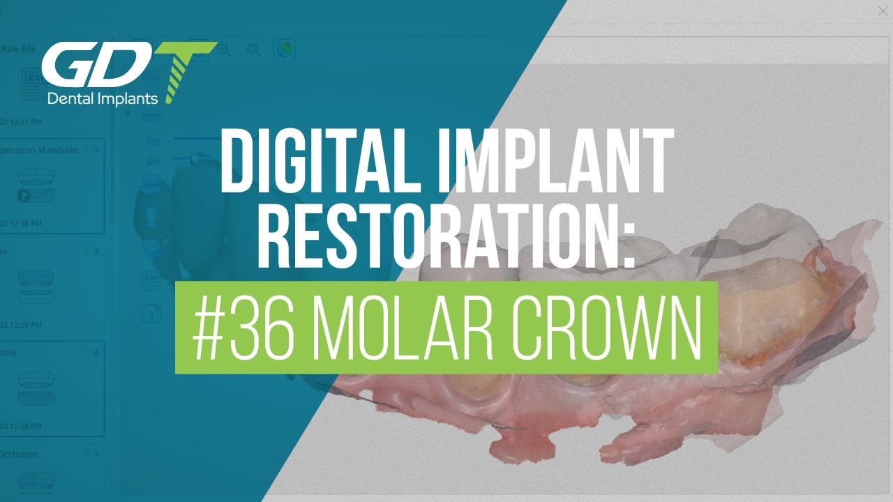
Digital Precision: Crown for #36 Molar with GDT Implants CAD/CAM and Advanced Tech
Get ready to witness the future of dental implant restoration!
Join Dr. Sarossi Benedek from Hungary as he takes us through the process of creating a single implant-based crown for the #36 molar using GDT Implant systems CAD/CAM digital library, 3Shape Trios3 Scanner, and Wieland Zenotec Select CNC.
Watch as the 3Shape Trios3 Scanner captures detailed 3D images of the patient's teeth and gums, creating a digital model of the patient's mouth that's used to plan the implant's placement and design the final restoration with precision.
See the crown come to life as it's fabricated using the Wieland Zenotec Select CNC, a computer numerical control machine that mills and fabricates dental restorations from a block of material, such as zirconia or titanium, using the digital design as a guide.
This cutting-edge digital workflow provides a high level of precision and accuracy, ensuring the best possible outcome for the patient.
{"description":"Introducing the ultimate emergency dental solution - GDT Supplies Temp Glue Over-The-Counter (OTC) Temporary Cement.\nDesigned for easy and convenient use, this pre-mixed dental cement is perfect for those unexpected moments when access to a dentist is not available.\nWith a fast cure time of 15 minutes, this dental cement provides an immediate solution for re-fixing crowns and bridges.\nAnd, with its two-stage setting, it allows for repositioning of the restoration within 3-5 minutes.\nAnd after 3 hours it's fully cured and provides an excellent and tight marginal seal.\nThis 7g container of dental cement is enough for 25+ repairs, and its hydrophilic properties make it easy to remove once professional help is available.\nTake the stress out of dental emergencies with GDT Supplies Temp Glue OTC Temporary Cement - manufactured for GDT Implants.","media_image":"https:\/\/i.ytimg.com\/vi\/1lLBcUKXNH8\/hq720.jpg","media_link":"https:\/\/www.youtube.com\/watch?v=1lLBcUKXNH8","media_page_content":{"type":"root","children":[{"type":"paragraph","children":[{"type":"text","value":"Introducing the ultimate emergency dental solution - GDT Supplies Temp Glue Over-The-Counter (OTC) Temporary Cement.\nDesigned for easy and convenient use, this pre-mixed dental cement is perfect for those unexpected moments when access to a dentist is not available.\nWith a fast cure time of 15 minutes, this dental cement provides an immediate solution for re-fixing crowns and bridges.\nAnd, with its two-stage setting, it allows for repositioning of the restoration within 3-5 minutes.\nAnd after 3 hours it's fully cured and provides an excellent and tight marginal seal.\nThis 7g container of dental cement is enough for 25+ repairs, and its hydrophilic properties make it easy to remove once professional help is available.\nTake the stress out of dental emergencies with GDT Supplies Temp Glue OTC Temporary Cement - manufactured for GDT Implants.\n\n"}]}]},"title":"GDT Temp Glue OTC: The Perfect Temporary Cement Solution"}
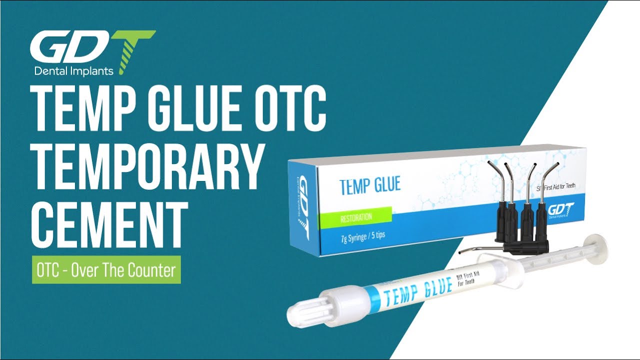
GDT Temp Glue OTC: The Perfect Temporary Cement Solution
Introducing the ultimate emergency dental solution - GDT Supplies Temp Glue Over-The-Counter (OTC) Temporary Cement.
Designed for easy and convenient use, this pre-mixed dental cement is perfect for those unexpected moments when access to a dentist is not available.
With a fast cure time of 15 minutes, this dental cement provides an immediate solution for re-fixing crowns and bridges.
And, with its two-stage setting, it allows for repositioning of the restoration within 3-5 minutes.
And after 3 hours it's fully cured and provides an excellent and tight marginal seal.
This 7g container of dental cement is enough for 25+ repairs, and its hydrophilic properties make it easy to remove once professional help is available.
Take the stress out of dental emergencies with GDT Supplies Temp Glue OTC Temporary Cement - manufactured for GDT Implants.
{"description":"Join us for the unboxing of our ABA Spiral Dental Implant, engineered for versatility across all surgical protocols, including immediate loading, two-stage, and flapless procedures.\n\nGDT ABA Implant features a tapered body with deep, aggressive apical threads, optimized for engagement in narrow ridges and poor bone conditions, without the need for bone grafting.\n\nMade from Titanium Grade 5 (Ti-6Al-4V ELI) and treated with SLA (Sandblast, Large Grit, Acid-etch) for improved osseointegration, this implant supports excellent primary stability and effective load distribution.\n\nIdeal for dentists seeking efficient handling and high compatibility across ridge types and indications.","media_image":"https:\/\/i.ytimg.com\/vi\/TpKGuEWZudU\/hqdefault.jpg","media_link":"https:\/\/www.youtube.com\/watch?v=TpKGuEWZudU","media_page_content":{"type":"root","children":[{"type":"paragraph","children":[{"type":"text","value":"Join us for the unboxing of our ABA Spiral Dental Implant, engineered for versatility across all surgical protocols, including immediate loading, two-stage, and flapless procedures.\n\nGDT ABA Implant features a tapered body with deep, aggressive apical threads, optimized for engagement in narrow ridges and poor bone conditions, without the need for bone grafting.\n\nMade from Titanium Grade 5 (Ti-6Al-4V ELI) and treated with SLA (Sandblast, Large Grit, Acid-etch) for improved osseointegration, this implant supports excellent primary stability and effective load distribution.\n\nIdeal for dentists seeking efficient handling and high compatibility across ridge types and indications.\n"}]}]},"title":"Unboxing MOR Spiral Implant | GDT Dental Implants"}
Unboxing MOR Spiral Implant | GDT Dental Implants
Join us for the unboxing of our ABA Spiral Dental Implant, engineered for versatility across all surgical protocols, including immediate loading, two-stage, and flapless procedures.
GDT ABA Implant features a tapered body with deep, aggressive apical threads, optimized for engagement in narrow ridges and poor bone conditions, without the need for bone grafting.
Made from Titanium Grade 5 (Ti-6Al-4V ELI) and treated with SLA (Sandblast, Large Grit, Acid-etch) for improved osseointegration, this implant supports excellent primary stability and effective load distribution.
Ideal for dentists seeking efficient handling and high compatibility across ridge types and indications.
{"description":"Introducing the GDT Implants Manual Torque Driver Handle - the ultimate tool for precision prosthetic implant placement.\nMade from durable stainless steel and this driver handle is engineered to provide accurate tightening of cover screws, healing caps, abutment screws, and overdenture attachments with a torque limitation of 15 Ncm.\nWith its 1.25mm driver hex, it fits with most leading implant brands, making it a versatile tool for any dental practice.\nThe driver handle also features a unique design that prevents over-tightening, ensuring optimal implant stability and patient safety.\nEffortlessly achieve precision and reliability with the GDT Implants Manual Torque Driver Handle.\nMade in Israel, it's easy to use and perfect for any dental professional looking to elevate their implant placement game.","media_image":"https:\/\/i.ytimg.com\/vi\/Q7NyEtWx_cE\/hqdefault.jpg","media_link":"https:\/\/www.youtube.com\/watch?v=Q7NyEtWx_cE","media_page_content":{"type":"root","children":[{"type":"paragraph","children":[{"type":"text","value":"Introducing the GDT Implants Manual Torque Driver Handle - the ultimate tool for precision prosthetic implant placement.\nMade from durable stainless steel and this driver handle is engineered to provide accurate tightening of cover screws, healing caps, abutment screws, and overdenture attachments with a torque limitation of 15 Ncm.\nWith its 1.25mm driver hex, it fits with most leading implant brands, making it a versatile tool for any dental practice.\nThe driver handle also features a unique design that prevents over-tightening, ensuring optimal implant stability and patient safety.\nEffortlessly achieve precision and reliability with the GDT Implants Manual Torque Driver Handle.\nMade in Israel, it's easy to use and perfect for any dental professional looking to elevate their implant placement game.\n\n"}]}]},"title":"GDT Manual Torque Driver Handle 1.25mm: The Future of Prosthetic Implant Placement"}

GDT Manual Torque Driver Handle 1.25mm: The Future of Prosthetic Implant Placement
Introducing the GDT Implants Manual Torque Driver Handle - the ultimate tool for precision prosthetic implant placement.
Made from durable stainless steel and this driver handle is engineered to provide accurate tightening of cover screws, healing caps, abutment screws, and overdenture attachments with a torque limitation of 15 Ncm.
With its 1.25mm driver hex, it fits with most leading implant brands, making it a versatile tool for any dental practice.
The driver handle also features a unique design that prevents over-tightening, ensuring optimal implant stability and patient safety.
Effortlessly achieve precision and reliability with the GDT Implants Manual Torque Driver Handle.
Made in Israel, it's easy to use and perfect for any dental professional looking to elevate their implant placement game.
{"description":"GDT Sponge Grafting Plug: The Future of Bone Grafting Procedures.\nRevolutionize your bone grafting procedures with GDT Sponge Grafting Plug - the ultimate one-stop solution for dental professionals.\nMade from a combination of pure bovine collagen and calcium phosphate hydroxyapatite, these sponge plugs offer a unique blend of bioactivity and ease of use.\nUnlike traditional particulate grafts, these plugs require no mixing and no membrane, making the grafting process quicker and more efficient.\nWith a diameter of Ø10mm and a length of 20mm, these sponge plugs are manufactured in Israel.\nGDT Sponge Grafting Plug offers a strong bond with bone as it resorbs, and releases calcium ions which create an environment favorable to rapid bone growth.\nThe material is radiolucent on the day of placement, becoming radiopaque 3-6 months later as it is replaced with native bone, enabling dental professionals to observe the healing process radiographically.\nSay goodbye to the hassle of traditional bone grafting procedures and experience the ease and efficiency of GDT Implants Sponge Grafting Plug.","media_image":"https:\/\/i.ytimg.com\/vi\/X2fUEceZh6U\/hqdefault.jpg","media_link":"https:\/\/www.youtube.com\/watch?v=X2fUEceZh6U","media_page_content":{"type":"root","children":[{"type":"paragraph","children":[{"type":"text","value":"GDT Sponge Grafting Plug: The Future of Bone Grafting Procedures.\nRevolutionize your bone grafting procedures with GDT Sponge Grafting Plug - the ultimate one-stop solution for dental professionals.\nMade from a combination of pure bovine collagen and calcium phosphate hydroxyapatite, these sponge plugs offer a unique blend of bioactivity and ease of use.\nUnlike traditional particulate grafts, these plugs require no mixing and no membrane, making the grafting process quicker and more efficient.\nWith a diameter of Ø10mm and a length of 20mm, these sponge plugs are manufactured in Israel.\nGDT Sponge Grafting Plug offers a strong bond with bone as it resorbs, and releases calcium ions which create an environment favorable to rapid bone growth.\nThe material is radiolucent on the day of placement, becoming radiopaque 3-6 months later as it is replaced with native bone, enabling dental professionals to observe the healing process radiographically.\nSay goodbye to the hassle of traditional bone grafting procedures and experience the ease and efficiency of GDT Implants Sponge Grafting Plug.\n\n"}]}]},"title":"GDT Sponge Grafting Plug: The Ultimate One-Stop Solution"}

GDT Sponge Grafting Plug: The Ultimate One-Stop Solution
GDT Sponge Grafting Plug: The Future of Bone Grafting Procedures.
Revolutionize your bone grafting procedures with GDT Sponge Grafting Plug - the ultimate one-stop solution for dental professionals.
Made from a combination of pure bovine collagen and calcium phosphate hydroxyapatite, these sponge plugs offer a unique blend of bioactivity and ease of use.
Unlike traditional particulate grafts, these plugs require no mixing and no membrane, making the grafting process quicker and more efficient.
With a diameter of Ø10mm and a length of 20mm, these sponge plugs are manufactured in Israel.
GDT Sponge Grafting Plug offers a strong bond with bone as it resorbs, and releases calcium ions which create an environment favorable to rapid bone growth.
The material is radiolucent on the day of placement, becoming radiopaque 3-6 months later as it is replaced with native bone, enabling dental professionals to observe the healing process radiographically.
Say goodbye to the hassle of traditional bone grafting procedures and experience the ease and efficiency of GDT Implants Sponge Grafting Plug.